 Movie
Movie Controller


+ Open data

- Basic information
| Entry | Database: PDB / ID: 5it4 | ||||||
|---|---|---|---|---|---|---|---|
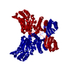
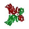
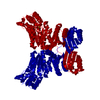
| Title | Crystal structure of Mycobacterium avium SerB2 mutant D343N | ||||||
 Components Components | Phosphoserine phosphatase | ||||||
 Keywords Keywords | HYDROLASE / HAD family / phosphoserine phosphatase / catalytic site mutant | ||||||
| Function / homology |  Function and homology information Function and homology informationphosphoserine phosphatase / L-phosphoserine phosphatase activity / L-serine biosynthetic process / magnesium ion binding / cytoplasm Similarity search - Function | ||||||
| Biological species |  Mycobacterium avium (bacteria) Mycobacterium avium (bacteria) | ||||||
| Method |  X-RAY DIFFRACTION / SYNCHROTRON / MOLECULAR REPLACEMENT / Resolution: 2.102 Å X-RAY DIFFRACTION / SYNCHROTRON / MOLECULAR REPLACEMENT / Resolution: 2.102 Å | ||||||
 Authors Authors | Shree, S. / Ramachandran, R. | ||||||
| Funding support |  India, 1items India, 1items
| ||||||
 Citation Citation | Journal: To be published Title: Crystal Structure of Mycobacterium avium SerB2 mutant D343N (MAV_3907) at pH 6.6 Authors: Shree, S. / Ramachandran, R. #1: Journal: J. Struct. Funct. Genomics / Year: 2011Title: SAD phasing using iodide ions in a high-throughput structural genomics environment. Authors: Abendroth, J. / Gardberg, A.S. / Robinson, J.I. / Christensen, J.S. / Staker, B.L. / Myler, P.J. / Stewart, L.J. / Edwards, T.E. | ||||||
| History |
|
- Structure visualization
| Structure viewer | Molecule: MolmilJmol/JSmol |
|---|
- Downloads & links
-Download
| PDBx/mmCIF format | 5it4.cif.gz | 165.8 KB | Display | PDBx/mmCIF format |
|---|---|---|---|---|
| PDB format | pdb5it4.ent.gz | 130.2 KB | Display | PDB format |
| PDBx/mmJSON format | 5it4.json.gz | Tree view | PDBx/mmJSON format | |
| Others |  Other downloads Other downloads |
-Validation report
| Summary document | 5it4_validation.pdf.gz | 424.2 KB | Display | wwPDB validaton report |
|---|---|---|---|---|
| Full document | 5it4_full_validation.pdf.gz | 426 KB | Display | |
| Data in XML | 5it4_validation.xml.gz | 16.7 KB | Display | |
| Data in CIF | 5it4_validation.cif.gz | 23.7 KB | Display | |
| Arichive directory | https://data.pdbj.org/pub/pdb/validation_reports/it/5it4ftp://data.pdbj.org/pub/pdb/validation_reports/it/5it4 | HTTPS FTP |
-Related structure data
| Related structure data |  3p96S S: Starting model for refinement |
|---|---|
| Similar structure data |


-Links
 PDBj
PDBj

- Assembly
| Deposited unit | 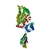
| ||||||||
|---|---|---|---|---|---|---|---|---|---|
| 1 | 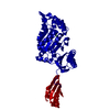
| ||||||||
| Unit cell |
|
-Components
| #1: Protein | Mass: 41786.836 Da / Num. of mol.: 1 / Fragment: UNP RESIDUES 5-400 / Mutation: G31R, D343N Source method: isolated from a genetically manipulated source Source: (gene. exp.) Mycobacterium avium (strain 104) (bacteria)Strain: 104 / Gene: serB, MAV_3907 / Production host: |
|---|---|
| #2: Chemical | ChemComp-MG /   Mass: 24.305 Da / Num. of mol.: 1 / Source method: obtained synthetically / Formula: Mg Mass: 24.305 Da / Num. of mol.: 1 / Source method: obtained synthetically / Formula: Mg |
| #3: Water | ChemComp-HOH /  Mass: 18.015 Da / Num. of mol.: 166 / Source method: isolated from a natural source / Formula: H2O Mass: 18.015 Da / Num. of mol.: 166 / Source method: isolated from a natural source / Formula: H2O |
-Experimental details
-Experiment
| Experiment | Method: X-RAY DIFFRACTION / Number of used crystals: 1 |
|---|
- Sample preparation
| Crystal | Density Matthews: 2.88 Å3/Da / Density % sol: 57.36 % |
|---|---|
| Crystal grow | Temperature: 293 K / Method: vapor diffusion, hanging drop / pH: 6.6 Details: 22- 23% PEG 8000, 0.1 M Magnesium acetate tetrahydrate, 0.1 M HEPES pH 6.6 |
-Data collection
| Diffraction | Mean temperature: 100 K |
|---|---|
| Diffraction source | Source: SYNCHROTRON / Site: ESRF  / Beamline: BM14 / Wavelength: 0.987 Å / Beamline: BM14 / Wavelength: 0.987 Å |
| Detector | Type: MARMOSAIC 225 mm CCD / Detector: CCD / Date: Dec 3, 2013 |
| Radiation | Protocol: SINGLE WAVELENGTH / Monochromatic (M) / Laue (L): M / Scattering type: x-ray |
| Radiation wavelength | Wavelength: 0.987 Å / Relative weight: 1 |
| Reflection | Resolution: 2.1→50 Å / Num. obs: 28166 / % possible obs: 98.6 % / Redundancy: 7.9 % / Net I/σ(I): 27.625 |
- Processing
| Software |
| |||||||||||||||||||||||||||||||||||||||||||||||||||||||||||||||||||||||||||||||||||||||||||||||||||||||||
|---|---|---|---|---|---|---|---|---|---|---|---|---|---|---|---|---|---|---|---|---|---|---|---|---|---|---|---|---|---|---|---|---|---|---|---|---|---|---|---|---|---|---|---|---|---|---|---|---|---|---|---|---|---|---|---|---|---|---|---|---|---|---|---|---|---|---|---|---|---|---|---|---|---|---|---|---|---|---|---|---|---|---|---|---|---|---|---|---|---|---|---|---|---|---|---|---|---|---|---|---|---|---|---|---|---|---|
| Refinement | Method to determine structure: MOLECULAR REPLACEMENT Starting model: 3P96 Resolution: 2.102→30.626 Å / SU ML: 0.24 / Cross valid method: NONE / σ(F): 1.35 / Phase error: 25.61 / Stereochemistry target values: ML
| |||||||||||||||||||||||||||||||||||||||||||||||||||||||||||||||||||||||||||||||||||||||||||||||||||||||||
| Solvent computation | Shrinkage radii: 0.9 Å / VDW probe radii: 1.11 Å / Solvent model: FLAT BULK SOLVENT MODEL | |||||||||||||||||||||||||||||||||||||||||||||||||||||||||||||||||||||||||||||||||||||||||||||||||||||||||
| Displacement parameters | Biso max: 106.82 Å2 / Biso mean: 47.5627 Å2 / Biso min: 27.27 Å2 | |||||||||||||||||||||||||||||||||||||||||||||||||||||||||||||||||||||||||||||||||||||||||||||||||||||||||
| Refinement step | Cycle: final / Resolution: 2.102→30.626 Å
| |||||||||||||||||||||||||||||||||||||||||||||||||||||||||||||||||||||||||||||||||||||||||||||||||||||||||
| Refine LS restraints |
| |||||||||||||||||||||||||||||||||||||||||||||||||||||||||||||||||||||||||||||||||||||||||||||||||||||||||
| LS refinement shell | Refine-ID: X-RAY DIFFRACTION / Total num. of bins used: 14
| |||||||||||||||||||||||||||||||||||||||||||||||||||||||||||||||||||||||||||||||||||||||||||||||||||||||||
| Refinement TLS params. | Method: refined / Origin x: -5.095 Å / Origin y: -10.7597 Å / Origin z: -38.967 Å
| |||||||||||||||||||||||||||||||||||||||||||||||||||||||||||||||||||||||||||||||||||||||||||||||||||||||||
| Refinement TLS group | Selection details: all |
