 Movie
Movie Controller

[English] 日本語
 Yorodumi
- PDB-5ipq: Cryo-EM structure of GluN1/GluN2B NMDA receptor in the DCKA/D-APV... -
+ Open data

- Basic information
| Entry | Database: PDB / ID: 5ipq | ||||||
|---|---|---|---|---|---|---|---|
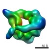
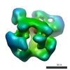
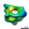
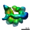
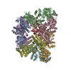
| Title | Cryo-EM structure of GluN1/GluN2B NMDA receptor in the DCKA/D-APV-bound conformation, state 2 | ||||||
 Components Components |
| ||||||
 Keywords Keywords | SIGNALING PROTEIN / ligand-gated ion channel / synaptic transmission | ||||||
| Function / homology |  Function and homology information Function and homology informationNMDA glutamate receptor activity / NMDA selective glutamate receptor complex / ligand-gated sodium channel activity / response to zinc ion / response to magnesium ion / glutamate-gated calcium ion channel activity / sodium ion transmembrane transport / ionotropic glutamate receptor signaling pathway / regulation of membrane potential / excitatory postsynaptic potential ...NMDA glutamate receptor activity / NMDA selective glutamate receptor complex / ligand-gated sodium channel activity / response to zinc ion / response to magnesium ion / glutamate-gated calcium ion channel activity / sodium ion transmembrane transport / ionotropic glutamate receptor signaling pathway / regulation of membrane potential / excitatory postsynaptic potential / transmitter-gated monoatomic ion channel activity involved in regulation of postsynaptic membrane potential / synaptic transmission, glutamatergic / postsynaptic density membrane / regulation of synaptic plasticity / calcium ion transmembrane transport / long-term synaptic potentiation / late endosome / signaling receptor activity / chemical synaptic transmission / postsynaptic membrane / lysosome / neuron projection / synapse / metal ion binding / plasma membrane Similarity search - Function | ||||||
| Biological species | |||||||
| Method | ELECTRON MICROSCOPY / single particle reconstruction / cryo EM / Resolution: 13.5 Å | ||||||
 Authors Authors | Zhu, S. / Stein, A.R. / Yoshioka, C. / Lee, C.H. / Goehring, A. / Mchaourab, S.H. / Gouaux, E. | ||||||
 Citation Citation | Journal: Cell / Year: 2016 Title: Mechanism of NMDA Receptor Inhibition and Activation. Authors: Shujia Zhu / Richard A Stein / Craig Yoshioka / Chia-Hsueh Lee / April Goehring / Hassane S Mchaourab / Eric Gouaux /  Abstract: N-methyl-D-aspartate receptors (NMDARs) are glutamate-gated, calcium-permeable ion channels that mediate synaptic transmission and underpin learning and memory. NMDAR dysfunction is directly ...N-methyl-D-aspartate receptors (NMDARs) are glutamate-gated, calcium-permeable ion channels that mediate synaptic transmission and underpin learning and memory. NMDAR dysfunction is directly implicated in diseases ranging from seizure to ischemia. Despite its fundamental importance, little is known about how the NMDAR transitions between inactive and active states and how small molecules inhibit or activate ion channel gating. Here, we report electron cryo-microscopy structures of the GluN1-GluN2B NMDA receptor in an ensemble of competitive antagonist-bound states, an agonist-bound form, and a state bound with agonists and the allosteric inhibitor Ro25-6981. Together with double electron-electron resonance experiments, we show how competitive antagonists rupture the ligand binding domain (LBD) gating "ring," how agonists retain the ring in a dimer-of-dimers configuration, and how allosteric inhibitors, acting within the amino terminal domain, further stabilize the LBD layer. These studies illuminate how the LBD gating ring is fundamental to signal transduction and gating in NMDARs. | ||||||
| History |
|
- Structure visualization
| Movie |
Movie viewer |
|---|---|
| Structure viewer | Molecule: MolmilJmol/JSmol |
- Downloads & links
-Download
| PDBx/mmCIF format | 5ipq.cif.gz | 374.2 KB | Display | PDBx/mmCIF format |
|---|---|---|---|---|
| PDB format | pdb5ipq.ent.gz | 229.1 KB | Display | PDB format |
| PDBx/mmJSON format | 5ipq.json.gz | Tree view | PDBx/mmJSON format | |
| Others |  Other downloads Other downloads |
-Validation report
| Summary document | 5ipq_validation.pdf.gz | 775.4 KB | Display | wwPDB validaton report |
|---|---|---|---|---|
| Full document | 5ipq_full_validation.pdf.gz | 776.9 KB | Display | |
| Data in XML | 5ipq_validation.xml.gz | 51.3 KB | Display | |
| Data in CIF | 5ipq_validation.cif.gz | 85.5 KB | Display | |
| Arichive directory | https://data.pdbj.org/pub/pdb/validation_reports/ip/5ipqftp://data.pdbj.org/pub/pdb/validation_reports/ip/5ipq | HTTPS FTP |
-Related structure data
| Related structure data | 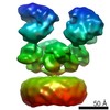 8101MC  8097C 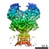 8098C  8102C 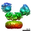 8103C 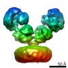 8104C 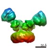 8105C  8106C  5iouC  5iovC  5iprC  5ipsC  5iptC 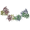 5ipuC  5ipvC M: map data used to model this data C: citing same article ( |
|---|---|
| Similar structure data |


-Links
 PDBj
PDBj

- Assembly
| Deposited unit | 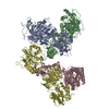
|
|---|---|
| 1 |
|
-Components
| #1: Protein | Mass: 92651.234 Da / Num. of mol.: 2 Mutation: K51F, R52F, N300Q, N350Q, N368D, N440D, N469D, K493A, K494A, E495A, G610R, I617L, D656R, N769E Source method: isolated from a genetically manipulated source Source: (gene. exp.)  Homo sapiens (human) / References: UniProt: C0KD18, UniProt: A0A1L8F5J9*PLUS Homo sapiens (human) / References: UniProt: C0KD18, UniProt: A0A1L8F5J9*PLUS#2: Protein | Mass: 93234.742 Da / Num. of mol.: 2 Mutation: M20S, G21R, C22A, A64E, N69Q, N343D, T490V, V615L, E654R, E655R Source method: isolated from a genetically manipulated source Source: (gene. exp.) Homo sapiens (human) / References: UniProt: A7XY94 |
|---|
-Experimental details
-Experiment
| Experiment | Method: ELECTRON MICROSCOPY |
|---|---|
| EM experiment | Aggregation state: PARTICLE / 3D reconstruction method: single particle reconstruction |
- Sample preparation
| Component | Name: GluN1-GluN2B receptor in the complex with competitive antagonists DCKA and D-APV Type: ORGANELLE OR CELLULAR COMPONENT / Entity ID: all / Source: RECOMBINANT |
|---|---|
| Source (natural) | Organism: |
| Source (recombinant) | Organism: Homo sapiens (human) / Cell: GnTI- / Plasmid: pEGBacmam |
| Buffer solution | pH: 6.5 |
| Specimen | Conc.: 4 mg/ml / Embedding applied: NO / Shadowing applied: NO / Staining applied: NO / Vitrification applied: YES |
| Vitrification | Instrument: FEI VITROBOT MARK III / Cryogen name: ETHANE / Humidity: 100 % / Chamber temperature: 18 K |
- Electron microscopy imaging
| Experimental equipment |  Model: Titan Krios / Image courtesy: FEI Company |
|---|---|
| Microscopy | Model: FEI TITAN KRIOS |
| Electron gun | Electron source:  FIELD EMISSION GUN / Accelerating voltage: 300 kV / Illumination mode: FLOOD BEAM FIELD EMISSION GUN / Accelerating voltage: 300 kV / Illumination mode: FLOOD BEAM |
| Electron lens | Mode: BRIGHT FIELD / Nominal defocus max: 3000 nm / Nominal defocus min: 1500 nm / Cs: 0.01 mm |
| Image recording | Electron dose: 10.5 e/Å2 / Detector mode: SUPER-RESOLUTION / Film or detector model: GATAN K2 SUMMIT (4k x 4k) |
- Processing
| EM software |
| ||||||||||||||||||||||||
|---|---|---|---|---|---|---|---|---|---|---|---|---|---|---|---|---|---|---|---|---|---|---|---|---|---|
| CTF correction | Type: PHASE FLIPPING AND AMPLITUDE CORRECTION | ||||||||||||||||||||||||
| Particle selection | Num. of particles selected: 393513 | ||||||||||||||||||||||||
| Symmetry | Point symmetry: C1 (asymmetric) | ||||||||||||||||||||||||
| 3D reconstruction | Resolution: 13.5 Å / Resolution method: FSC 0.143 CUT-OFF / Num. of particles: 38007 / Symmetry type: POINT |
