 Movie
Movie Controller

[English] 日本語
 Yorodumi
- EMDB-1438: Architecture of the yeast Rrp44 exosome complex suggests routes o... -
+ Open data

- Basic information
| Entry | Database: EMDB / ID: EMD-1438 | |||||||||
|---|---|---|---|---|---|---|---|---|---|---|
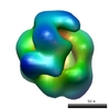
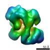
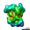
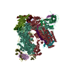
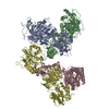
| Title | Architecture of the yeast Rrp44 exosome complex suggests routes of RNA recruitment for 3' end processing. | |||||||||
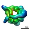
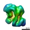
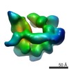
 Map data Map data | This is the map of yeast Rrp44-associated exosome complex | |||||||||
 Sample Sample |
| |||||||||
| Biological species |  | |||||||||
| Method | single particle reconstruction / cryo EM / negative staining / Resolution: 19.0 Å | |||||||||
 Authors Authors | Wang H-W / Wang J / Ding F / Callahan K / Bratkowski MA / Butler JS / Nogales E / Ke A | |||||||||
 Citation Citation | Journal: Proc Natl Acad Sci U S A / Year: 2007 Title: Architecture of the yeast Rrp44 exosome complex suggests routes of RNA recruitment for 3' end processing. Authors: Hong-Wei Wang / Jianjun Wang / Fang Ding / Kevin Callahan / Matthew A Bratkowski / J Scott Butler / Eva Nogales / Ailong Ke /  Abstract: The eukaryotic core exosome (CE) is a conserved nine-subunit protein complex important for 3' end trimming and degradation of RNA. In yeast, the Rrp44 protein constitutively associates with the CE ...The eukaryotic core exosome (CE) is a conserved nine-subunit protein complex important for 3' end trimming and degradation of RNA. In yeast, the Rrp44 protein constitutively associates with the CE and provides the sole source of processive 3'-to-5' exoribonuclease activity. Here we present EM reconstructions of the core and Rrp44-bound exosome complexes. The two-lobed Rrp44 protein binds to the RNase PH domain side of the exosome and buttresses the bottom of the exosome-processing chamber. The Rrp44 C-terminal body part containing an RNase II-type active site is anchored to the exosome through a conserved set of interactions mainly to the Rrp45 and Rrp43 subunit, whereas the Rrp44 N-terminal head part is anchored to the Rrp41 subunit and may function as a roadblock to restrict access of RNA to the active site in the body region. The Rrp44-exosome (RE) architecture suggests an active site sequestration mechanism for strict control of 3' exoribonuclease activity in the RE complex. | |||||||||
| History |
|
- Structure visualization
| Movie |
 Movie viewer Movie viewer |
|---|---|
| Structure viewer | EM map: SurfViewMolmilJmol/JSmol |
| Supplemental images |
 UCSF Chimera
- Downloads & links
-EMDB archive
| Map data | emd_1438.map.gz | 1.2 MB | EMDB map data format | |
|---|---|---|---|---|
| Header (meta data) | emd-1438-v30.xmlemd-1438.xml | 17.3 KB 17.3 KB | Display Display | EMDB header |
| Images |  1438.gif 1438.gif | 62.8 KB | ||
| Archive directory |  http://ftp.pdbj.org/pub/emdb/structures/EMD-1438ftp://ftp.pdbj.org/pub/emdb/structures/EMD-1438 http://ftp.pdbj.org/pub/emdb/structures/EMD-1438ftp://ftp.pdbj.org/pub/emdb/structures/EMD-1438 | HTTPS FTP |
-Related structure data
-Links
| EMDB pages | EMDB (EBI/PDBe) / EMDataResource |
|---|
-Map
| File | Download / File: emd_1438.map.gz / Format: CCP4 / Size: 1.4 MB / Type: IMAGE STORED AS FLOATING POINT NUMBER (4 BYTES) | ||||||||||||||||||||||||||||||||||||||||||||||||||||||||||||||||||||
|---|---|---|---|---|---|---|---|---|---|---|---|---|---|---|---|---|---|---|---|---|---|---|---|---|---|---|---|---|---|---|---|---|---|---|---|---|---|---|---|---|---|---|---|---|---|---|---|---|---|---|---|---|---|---|---|---|---|---|---|---|---|---|---|---|---|---|---|---|---|
| Annotation | This is the map of yeast Rrp44-associated exosome complex | ||||||||||||||||||||||||||||||||||||||||||||||||||||||||||||||||||||
| Projections & slices | Image control
Images are generated by Spider. | ||||||||||||||||||||||||||||||||||||||||||||||||||||||||||||||||||||
| Voxel size | X=Y=Z: 5.18 Å | ||||||||||||||||||||||||||||||||||||||||||||||||||||||||||||||||||||
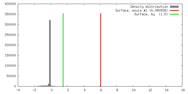
| Density |
| ||||||||||||||||||||||||||||||||||||||||||||||||||||||||||||||||||||
| Symmetry | Space group: 1 | ||||||||||||||||||||||||||||||||||||||||||||||||||||||||||||||||||||
| Details | EMDB XML:
CCP4 map header:
| ||||||||||||||||||||||||||||||||||||||||||||||||||||||||||||||||||||
 Z (Sec.)
 Y (Row.)
 X (Col.)


-Supplemental data
- Sample components
+Entire : Rrp44-associated exosome complex
+Supramolecule #1000: Rrp44-associated exosome complex
+Macromolecule #1: Rrp44
+Macromolecule #2: Rrp43
+Macromolecule #3: Rrp4
+Macromolecule #4: Csl4
+Macromolecule #5: Rrp45
+Macromolecule #6: Rrp46-TAP
+Macromolecule #7: Rrp41
+Macromolecule #8: Rrp42
+Macromolecule #9: Mtr3
+Macromolecule #10: Rrp40
-Experimental details
-Structure determination
| Method | negative staining, cryo EM |
|---|---|
 Processing Processing | single particle reconstruction |
| Aggregation state | particle |
-Sample preparation
| Concentration | 0.04 mg/mL |
|---|---|
| Buffer | pH: 7.5 Details: 25mM Tris-HCl, 50 mM NaCl, 2 mM DTT, and 10 uM ZnCl2 |
| Staining | Type: NEGATIVE Details: Four microliters of the protein solution was negatively stained with 2% uranyl formate solution between two thin layers of carbon on a copper grid by using the sandwich method |
| Grid | Details: 400 mesh copper grid |
| Vitrification | Cryogen name: ETHANE |
- Electron microscopy
| Microscope | FEI TECNAI 12 |
|---|---|
| Temperature | Average: 300 K |
| Alignment procedure | Legacy - Astigmatism: objective lens astigmatism was corrected at |
| Details | Low dose mode for taking pictures |
| Date | Aug 1, 2006 |
| Image recording | Category: FILM / Film or detector model: KODAK SO-163 FILM / Digitization - Scanner: OTHER / Digitization - Sampling interval: 12.7 µm / Number real images: 50 / Average electron dose: 20 e/Å2 / Details: The scanner was Nikon Super Coolscan 8000 / Od range: 1.4 / Bits/pixel: 14 |
| Tilt angle min | 0 |
| Electron beam | Acceleration voltage: 120 kV / Electron source: LAB6 |
| Electron optics | Calibrated magnification: 49000 / Illumination mode: FLOOD BEAM / Imaging mode: BRIGHT FIELD / Cs: 6.6 mm / Nominal defocus max: 0.9 µm / Nominal defocus min: 0.7 µm / Nominal magnification: 49000 |
| Sample stage | Specimen holder: normal single-tilt holder / Specimen holder model: OTHER / Tilt angle max: 55 |
-Image processing
| Final reconstruction | Applied symmetry - Point group: C1 (asymmetric) / Algorithm: OTHER / Resolution.type: BY AUTHOR / Resolution: 19.0 Å / Resolution method: FSC 0.5 CUT-OFF / Software - Name: IMAGIC, SPIDER Details: Final map were calculated from all the untilted particles Number images used: 3020 |
|---|---|
| Final angle assignment | Details: SPIDER: theta 90 degrees, phi 90 degrees |
| Final two d classification | Number classes: 50 |
-Atomic model buiding 1
| Software | Name: Situs |
|---|---|
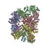
| Details | Protocol: Rigid Body. 2NN6.pdb and 2IX0.pdb were used to dock in the map. The latter one was separated to three domains after the automatic docking and manually adjusted to fit in the map locally in Chimera. |
| Refinement | Protocol: RIGID BODY FIT |
