 Movie
Movie Controller


+ Open data

- Basic information
| Entry | Database: PDB / ID: 4u39 | ||||||
|---|---|---|---|---|---|---|---|
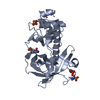
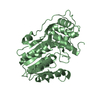
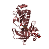
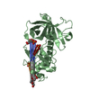
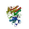
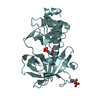
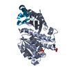
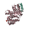
| Title | Crystal Structure of FtsZ:MciZ Complex from Bacillus subtilis | ||||||
 Components Components |
| ||||||
 Keywords Keywords | CELL CYCLE / FtsZ / MciZ / protein complex | ||||||
| Function / homology |  Function and homology information Function and homology informationcell septum / division septum assembly / FtsZ-dependent cytokinesis / sporulation resulting in formation of a cellular spore / cell division site / protein polymerization / cell division / GTPase activity / GTP binding / identical protein binding / cytoplasm Similarity search - Function | ||||||
| Biological species |  | ||||||
| Method |  X-RAY DIFFRACTION / SYNCHROTRON / MOLECULAR REPLACEMENT / Resolution: 3.194 Å X-RAY DIFFRACTION / SYNCHROTRON / MOLECULAR REPLACEMENT / Resolution: 3.194 Å | ||||||
 Authors Authors | Bisson-Filho, A.W. / Discola, K.F. / Castellen, P. / Blasios, V. / Martins, A. / Sforca, M.L. / Garcia, W. / Zeri, A.C. / Erickson, H.P. / Dessen, A. / Gueiros-Filho, F.J. | ||||||
 Citation Citation | Journal: To be Published Title: Crystal Structure of FtsZ:MciZ Complex from Bacillus subtilis Authors: Bisson-Filho, A.W. / Discola, K.F. / Castellen, P. / Blasios, V. / Martins, A. / Sforca, M.L. / Garcia, W. / Zeri, A.C. / Erickson, H.P. / Dessen, A. / Gueiros-Filho, F.J. | ||||||
| History |
|
- Structure visualization
| Structure viewer | Molecule: MolmilJmol/JSmol |
|---|
- Downloads & links
-Download
| PDBx/mmCIF format | 4u39.cif.gz | 491.2 KB | Display | PDBx/mmCIF format |
|---|---|---|---|---|
| PDB format | pdb4u39.ent.gz | 394.7 KB | Display | PDB format |
| PDBx/mmJSON format | 4u39.json.gz | Tree view | PDBx/mmJSON format | |
| Others |  Other downloads Other downloads |
-Validation report
| Arichive directory | https://data.pdbj.org/pub/pdb/validation_reports/u3/4u39ftp://data.pdbj.org/pub/pdb/validation_reports/u3/4u39 | HTTPS FTP |
|---|
-Related structure data
| Related structure data |  2vxyS S: Starting model for refinement |
|---|---|
| Similar structure data |


-Links
 PDBj
PDBj


- Assembly


-Components
| #1: Protein | Mass: 31476.879 Da / Num. of mol.: 9 / Fragment: UNP residues 12-315 Source method: isolated from a genetically manipulated source Source: (gene. exp.) #2: Protein | Mass: 6957.148 Da / Num. of mol.: 9 Source method: isolated from a genetically manipulated source Source: (gene. exp.) #3: Chemical | ChemComp-PO4 /   Mass: 94.971 Da / Num. of mol.: 16 / Source method: obtained synthetically / Formula: PO4 Mass: 94.971 Da / Num. of mol.: 16 / Source method: obtained synthetically / Formula: PO4 |
|---|
-Experimental details
-Experiment
| Experiment | Method: X-RAY DIFFRACTION |
|---|
- Sample preparation
| Crystal | Density Matthews: 3.09 Å3/Da / Density % sol: 60.18 % |
|---|---|
| Crystal grow | Temperature: 293 K / Method: vapor diffusion, hanging drop / pH: 7 / Details: 1.6 M sodium/potassium phosphate, pH 7.0 |
-Data collection
| Diffraction | Mean temperature: 100 K |
|---|---|
| Diffraction source | Source: SYNCHROTRON / Site: ESRF  / Beamline: ID14-4 / Wavelength: 1.0044 Å / Beamline: ID14-4 / Wavelength: 1.0044 Å |
| Detector | Type: ADSC QUANTUM 315r / Detector: CCD / Date: Feb 5, 2013 |
| Radiation | Monochromator: Si(111) / Protocol: SINGLE WAVELENGTH / Monochromatic (M) / Laue (L): M / Scattering type: x-ray |
| Radiation wavelength | Wavelength: 1.0044 Å / Relative weight: 1 |
| Reflection | Resolution: 3.19→46.593 Å / Num. obs: 72155 / % possible obs: 97.5 % / Redundancy: 17.33 % / Rsym value: 0.058 / Net I/σ(I): 62.64 |
| Reflection shell | Resolution: 3.19→3.39 Å / Rmerge(I) obs: 0.37 / Mean I/σ(I) obs: 12.7 / % possible all: 91.2 |
- Processing
| Software | Name: PHENIX / Version: (phenix.refine: 1.8_1069) / Classification: refinement | |||||||||||||||||||||||||||||||||||||||||||||||||||||||||||||||||||||||||||||||||||||||||||||||||||||||||||||||||||||||||||||||||||||||||||||||||||||||||||||||||||||||||||||||||||||||||||||
|---|---|---|---|---|---|---|---|---|---|---|---|---|---|---|---|---|---|---|---|---|---|---|---|---|---|---|---|---|---|---|---|---|---|---|---|---|---|---|---|---|---|---|---|---|---|---|---|---|---|---|---|---|---|---|---|---|---|---|---|---|---|---|---|---|---|---|---|---|---|---|---|---|---|---|---|---|---|---|---|---|---|---|---|---|---|---|---|---|---|---|---|---|---|---|---|---|---|---|---|---|---|---|---|---|---|---|---|---|---|---|---|---|---|---|---|---|---|---|---|---|---|---|---|---|---|---|---|---|---|---|---|---|---|---|---|---|---|---|---|---|---|---|---|---|---|---|---|---|---|---|---|---|---|---|---|---|---|---|---|---|---|---|---|---|---|---|---|---|---|---|---|---|---|---|---|---|---|---|---|---|---|---|---|---|---|---|---|---|---|---|
| Refinement | Method to determine structure: MOLECULAR REPLACEMENT Starting model: PDB ENTRY 2VXY Resolution: 3.194→46.593 Å / SU ML: 0.45 / Cross valid method: FREE R-VALUE / σ(F): 1.99 / Phase error: 29.24 / Stereochemistry target values: ML
| |||||||||||||||||||||||||||||||||||||||||||||||||||||||||||||||||||||||||||||||||||||||||||||||||||||||||||||||||||||||||||||||||||||||||||||||||||||||||||||||||||||||||||||||||||||||||||||
| Solvent computation | Shrinkage radii: 0.9 Å / VDW probe radii: 1.11 Å / Solvent model: FLAT BULK SOLVENT MODEL | |||||||||||||||||||||||||||||||||||||||||||||||||||||||||||||||||||||||||||||||||||||||||||||||||||||||||||||||||||||||||||||||||||||||||||||||||||||||||||||||||||||||||||||||||||||||||||||
| Refinement step | Cycle: LAST / Resolution: 3.194→46.593 Å
| |||||||||||||||||||||||||||||||||||||||||||||||||||||||||||||||||||||||||||||||||||||||||||||||||||||||||||||||||||||||||||||||||||||||||||||||||||||||||||||||||||||||||||||||||||||||||||||
| Refine LS restraints |
| |||||||||||||||||||||||||||||||||||||||||||||||||||||||||||||||||||||||||||||||||||||||||||||||||||||||||||||||||||||||||||||||||||||||||||||||||||||||||||||||||||||||||||||||||||||||||||||
| LS refinement shell |
|
