 Movie
Movie Controller

[English] 日本語
 Yorodumi
- PDB-5h3r: Crystal Structure of mutant MarR C80S from E.coli complexed with ... -
+ Open data

- Basic information
| Entry | Database: PDB / ID: 5h3r | ||||||
|---|---|---|---|---|---|---|---|
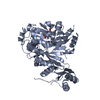
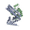
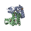
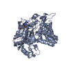
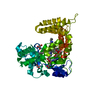
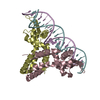
| Title | Crystal Structure of mutant MarR C80S from E.coli complexed with operator DNA | ||||||
 Components Components |
| ||||||
 Keywords Keywords | TRANSCRIPTION/DNA / MarR family protein / HTH motif / protein-DNA complex / Transcription Factor / TRANSCRIPTION-DNA complex | ||||||
| Function / homology |  Function and homology information Function and homology informationcellular response to antibiotic / response to stress / response to heat / DNA-binding transcription factor activity / negative regulation of DNA-templated transcription / regulation of DNA-templated transcription / DNA binding Similarity search - Function | ||||||
| Biological species |  synthetic construct (others) | ||||||
| Method |  X-RAY DIFFRACTION / SYNCHROTRON / MOLECULAR REPLACEMENT / Resolution: 2.67 Å X-RAY DIFFRACTION / SYNCHROTRON / MOLECULAR REPLACEMENT / Resolution: 2.67 Å | ||||||
 Authors Authors | Zhu, R. / Lou, H. / Hao, Z. | ||||||
 Citation Citation | Journal: J. Biol. Inorg. Chem. / Year: 2017 Title: Structural characterization of the DNA-binding mechanism underlying the copper(II)-sensing MarR transcriptional regulator. Authors: Zhu, R. / Hao, Z. / Lou, H. / Song, Y. / Zhao, J. / Chen, Y. / Zhu, J. / Chen, P.R. | ||||||
| History |
|
- Structure visualization
| Structure viewer | Molecule: MolmilJmol/JSmol |
|---|
- Downloads & links
-Download
| PDBx/mmCIF format | 5h3r.cif.gz | 92 KB | Display | PDBx/mmCIF format |
|---|---|---|---|---|
| PDB format | pdb5h3r.ent.gz | 65.5 KB | Display | PDB format |
| PDBx/mmJSON format | 5h3r.json.gz | Tree view | PDBx/mmJSON format | |
| Others |  Other downloads Other downloads |
-Validation report
| Arichive directory | https://data.pdbj.org/pub/pdb/validation_reports/h3/5h3rftp://data.pdbj.org/pub/pdb/validation_reports/h3/5h3r | HTTPS FTP |
|---|
-Related structure data
| Related structure data |  3q5fS S: Starting model for refinement |
|---|---|
| Similar structure data |


-Links
 PDBj
PDBj


- Assembly
| Deposited unit | 
| ||||||||
|---|---|---|---|---|---|---|---|---|---|
| 1 |
| ||||||||
| Unit cell |
|
-Components
| #1: DNA chain | Mass: 6413.159 Da / Num. of mol.: 1 / Source method: obtained synthetically / Source: (synth.) synthetic construct (others) | ||
|---|---|---|---|
| #2: DNA chain | Mass: 6471.211 Da / Num. of mol.: 1 / Source method: obtained synthetically / Source: (synth.) synthetic construct (others) | ||
| #3: Protein | Mass: 16353.234 Da / Num. of mol.: 2 / Mutation: C80S Source method: isolated from a genetically manipulated source Source: (gene. exp.) #4: Water | ChemComp-HOH / |  Mass: 18.015 Da / Num. of mol.: 24 / Source method: isolated from a natural source / Formula: H2O Mass: 18.015 Da / Num. of mol.: 24 / Source method: isolated from a natural source / Formula: H2O |
-Experimental details
-Experiment
| Experiment | Method: X-RAY DIFFRACTION / Number of used crystals: 1 |
|---|
- Sample preparation
| Crystal | Density Matthews: 3.14 Å3/Da / Density % sol: 60.83 % |
|---|---|
| Crystal grow | Temperature: 293 K / Method: vapor diffusion, sitting drop Details: PEG 5000 MME, sodium cacodylate, magnesium chloride, sodium chloride PH range: 5.6-6.2 |
-Data collection
| Diffraction | Mean temperature: 100 K |
|---|---|
| Diffraction source | Source: SYNCHROTRON / Site: SSRF  / Beamline: BL17U / Wavelength: 1.0055 Å / Beamline: BL17U / Wavelength: 1.0055 Å |
| Detector | Type: ADSC QUANTUM 315r / Detector: CCD / Date: Oct 10, 2014 |
| Radiation | Protocol: SINGLE WAVELENGTH / Monochromatic (M) / Laue (L): M / Scattering type: x-ray |
| Radiation wavelength | Wavelength: 1.0055 Å / Relative weight: 1 |
| Reflection | Resolution: 2.67→50 Å / Num. all: 16064 / Num. obs: 16057 / % possible obs: 100 % / Observed criterion σ(F): 0 / Observed criterion σ(I): -3 / Redundancy: 9.5 % / Biso Wilson estimate: 55.4 Å2 / Rmerge(I) obs: 0.112 / Rsym value: 0.112 / Net I/σ(I): 24.03 |
| Reflection shell | Resolution: 2.67→2.72 Å / Redundancy: 9.6 % / Rmerge(I) obs: 0.986 / Mean I/σ(I) obs: 3 / % possible all: 100 |
- Processing
| Software |
| |||||||||||||||||||||||||||||||||||||||||||||||||||||||||||||||||||||||||||||
|---|---|---|---|---|---|---|---|---|---|---|---|---|---|---|---|---|---|---|---|---|---|---|---|---|---|---|---|---|---|---|---|---|---|---|---|---|---|---|---|---|---|---|---|---|---|---|---|---|---|---|---|---|---|---|---|---|---|---|---|---|---|---|---|---|---|---|---|---|---|---|---|---|---|---|---|---|---|---|
| Refinement | Method to determine structure: MOLECULAR REPLACEMENT Starting model: 3Q5F Resolution: 2.67→33.717 Å / SU ML: 0.42 / Cross valid method: FREE R-VALUE / σ(F): 1.39 / Phase error: 28.88 / Stereochemistry target values: ML
| |||||||||||||||||||||||||||||||||||||||||||||||||||||||||||||||||||||||||||||
| Solvent computation | Shrinkage radii: 0.9 Å / VDW probe radii: 1.11 Å / Solvent model: FLAT BULK SOLVENT MODEL | |||||||||||||||||||||||||||||||||||||||||||||||||||||||||||||||||||||||||||||
| Displacement parameters | Biso mean: 60.2 Å2 | |||||||||||||||||||||||||||||||||||||||||||||||||||||||||||||||||||||||||||||
| Refinement step | Cycle: LAST / Resolution: 2.67→33.717 Å
| |||||||||||||||||||||||||||||||||||||||||||||||||||||||||||||||||||||||||||||
| Refine LS restraints |
| |||||||||||||||||||||||||||||||||||||||||||||||||||||||||||||||||||||||||||||
| LS refinement shell |
|
