[English] 日本語
Yorodumi
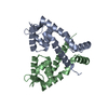
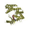
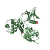
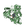
- PDB-3vod: Crystal Structure of mutant MarR C80S from E.coli -

+
Open data


ID or keywords:

Loading...

-
Basic information

Entry
Database: PDB / ID: 3vod
TitleCrystal Structure of mutant MarR C80S from E.coli
ComponentsMultiple antibiotic resistance protein marRMultiple drug resistance
KeywordsTRANSCRIPTION / winged helix-turn-helix DNA binding motif
Function / homology
Function and homology information


cellular response to antibiotic / response to heat / DNA-binding transcription factor activity / negative regulation of DNA-templated transcription / regulation of DNA-templated transcription / DNA binding
Similarity search - Function
Transcriptional regulator MarR-type, conserved site / MarR-type HTH domain signature. / MarR family / MarR-type HTH domain profile. / helix_turn_helix multiple antibiotic resistance protein / MarR-type HTH domain / Winged helix-like DNA-binding domain superfamily/Winged helix DNA-binding domain / Arc Repressor Mutant, subunit A / Winged helix DNA-binding domain superfamily / Winged helix-like DNA-binding domain superfamily ...Transcriptional regulator MarR-type, conserved site / MarR-type HTH domain signature. / MarR family / MarR-type HTH domain profile. / helix_turn_helix multiple antibiotic resistance protein / MarR-type HTH domain / Winged helix-like DNA-binding domain superfamily/Winged helix DNA-binding domain / Arc Repressor Mutant, subunit A / Winged helix DNA-binding domain superfamily / Winged helix-like DNA-binding domain superfamily / Orthogonal Bundle / Mainly Alpha
Similarity search - Domain/homology
Multiple antibiotic resistance protein MarR
Similarity search - Component
Biological speciesEscherichia coli (E. coli)
MethodX-RAY DIFFRACTION / SYNCHROTRON / MOLECULAR REPLACEMENT / molecular replacement / Resolution: 2.6 Å
AuthorsLou, H. / Zhu, R. / Hao, Z.
CitationJournal: Nat.Chem.Biol. / Year: 2014
Title: The multiple antibiotic resistance regulator MarR is a copper sensor in Escherichia coli.
Authors: Hao, Z. / Lou, H. / Zhu, R. / Zhu, J. / Zhang, D. / Zhao, B.S. / Zeng, S. / Chen, X. / Chan, J. / He, C. / Chen, P.R.
History
DepositionJan 21, 2012Deposition site: PDBJ / Processing site: PDBJ
Revision 1.0Mar 6, 2013Provider: repository / Type: Initial release
Revision 1.1Apr 9, 2014Group: Database references
Revision 1.2Nov 22, 2017Group: Refinement description / Category: software
Item: _software.classification / _software.contact_author ..._software.classification / _software.contact_author / _software.contact_author_email / _software.date / _software.language / _software.location / _software.name / _software.type / _software.version
Revision 1.3Mar 20, 2024Group: Data collection / Database references / Refinement description
Category: chem_comp_atom / chem_comp_bond ...chem_comp_atom / chem_comp_bond / database_2 / struct_ncs_dom_lim / struct_ref_seq_dif
Item: _database_2.pdbx_DOI / _database_2.pdbx_database_accession ..._database_2.pdbx_DOI / _database_2.pdbx_database_accession / _struct_ncs_dom_lim.beg_auth_comp_id / _struct_ncs_dom_lim.beg_label_asym_id / _struct_ncs_dom_lim.beg_label_comp_id / _struct_ncs_dom_lim.beg_label_seq_id / _struct_ncs_dom_lim.end_auth_comp_id / _struct_ncs_dom_lim.end_label_asym_id / _struct_ncs_dom_lim.end_label_comp_id / _struct_ncs_dom_lim.end_label_seq_id / _struct_ref_seq_dif.details

-
Structure visualization

Structure viewerMolecule:
MolmilJmol/JSmol

Downloads & links

-
Assembly

Deposited unit
A: Multiple antibiotic resistance protein marR
B: Multiple antibiotic resistance protein marR


Theoretical massNumber of molelcules
Total (without water)32,1422
Polymers32,1422
Non-polymers00
Water0
1


  • Idetical with deposited unit
  • defined by author&software
TypeNameSymmetry operationNumber
identity operation1_555x,y,z1
Buried area3850 Å2
ΔGint-28 kcal/mol
Surface area15310 Å2
MethodPISA
Unit cell
Length a, b, c (Å)62.162, 62.162, 131.466
Angle α, β, γ (deg.)90.000, 90.000, 90.000
Int Tables number96
Space group name H-MP43212
Noncrystallographic symmetry (NCS)NCS domain:
IDEns-IDDetails
11A
21B

NCS domain segments:

Component-ID: 1 / Ens-ID: 1 / Beg auth comp-ID: ILE / Beg label comp-ID: ILE / End auth comp-ID: PRO / End label comp-ID: PRO / Refine code: 4 / Auth seq-ID: 11 - 144 / Label seq-ID: 11 - 144

Dom-IDAuth asym-IDLabel asym-ID
1AA
2BB

-
Components

#1: Protein Multiple antibiotic resistance protein marR / Multiple drug resistance


Mass: 16070.957 Da / Num. of mol.: 2 / Mutation: C80S
Source method: isolated from a genetically manipulated source
Source: (gene. exp.) Escherichia coli (E. coli) / Strain: K12 / Gene: b1530, cfxB, inaR, JW5248, marR, soxQ / Plasmid: pET15b / Production host: Escherichia coli (E. coli) / Strain (production host): BL21(DE3) / References: UniProt: P27245

-
Experimental details

-
Experiment

ExperimentMethod: X-RAY DIFFRACTION / Number of used crystals: 1

-
Sample preparation

CrystalDensity Matthews: 1.98 Å3/Da / Density % sol: 37.74 % / Mosaicity: 0.817 °
Crystal growTemperature: 293 K / Method: vapor diffusion, sitting drop / pH: 7.6
Details: 0.03M Citric Acid, 0.07M BIS-TRIS, pH7.6, 24% PEG 3350,0.3M NaCl, 10mM DTT, vapor diffusion, sitting drop, temperature 293K

-
Data collection

DiffractionMean temperature: 100 K
Diffraction sourceSource: SYNCHROTRON / Site: BSRF / Beamline: 3W1A / Wavelength: 1 Å
DetectorDate: Jul 11, 2011
RadiationProtocol: SINGLE WAVELENGTH / Monochromatic (M) / Laue (L): M / Scattering type: x-ray
Radiation wavelengthWavelength: 1 Å / Relative weight: 1
ReflectionResolution: 2.6→40 Å / Num. obs: 8479 / % possible obs: 99.8 % / Redundancy: 6 % / Rmerge(I) obs: 0.072 / Χ2: 1.904 / Net I/σ(I): 16.3
Reflection shell
Resolution (Å)Redundancy (%)Rmerge(I) obsNum. unique allΧ2Diffraction-ID% possible all
2.6-2.696.20.3868220.9651100
2.69-2.86.10.2778190.9661100
2.8-2.936.10.2158261.0141100
2.93-3.086.20.1568221.1981100
3.08-3.2860.1048381.361100
3.28-3.536.20.0778281.731100
3.53-3.8860.0688532.2231100
3.88-4.4460.068602.815199.8
4.44-5.595.90.0558683.115199.7
5.59-305.40.0649433.692198.5

-
Phasing

PhasingMethod: molecular replacement
Phasing MRModel details: Phaser MODE: MR_AUTO
Highest resolutionLowest resolution
Rotation2.6 Å29.06 Å
Translation2.6 Å29.06 Å

-
Processing

Software
NameVersionClassificationNB
DENZOdata reduction
SCALEPACKdata scaling
PHASER2.3.0phasing
REFMAC5.6.0117refinement
PDB_EXTRACT3.1data extraction
RefinementMethod to determine structure: MOLECULAR REPLACEMENT / Resolution: 2.6→30 Å / Cor.coef. Fo:Fc: 0.934 / Cor.coef. Fo:Fc free: 0.911 / Occupancy max: 1 / Occupancy min: 1 / SU B: 40.259 / SU ML: 0.358 / Cross valid method: THROUGHOUT / σ(F): 0 / ESU R Free: 0.383 / Stereochemistry target values: MAXIMUM LIKELIHOOD
Details: HYDROGENS HAVE BEEN USED IF PRESENT IN THE INPUT U VALUES
RfactorNum. reflection% reflectionSelection details
Rfree0.2809 390 4.7 %RANDOM
Rwork0.2557 ---
obs0.2568 8380 99.02 %-
Solvent computationIon probe radii: 0.8 Å / Shrinkage radii: 0.8 Å / VDW probe radii: 1.2 Å / Solvent model: BABINET MODEL WITH MASK
Displacement parametersBiso max: 174.68 Å2 / Biso mean: 80.7712 Å2 / Biso min: 44.78 Å2
Baniso -1Baniso -2Baniso -3
1-0.17 Å20 Å20 Å2
2--0.17 Å20 Å2
3----0.34 Å2
Refinement stepCycle: LAST / Resolution: 2.6→30 Å
ProteinNucleic acidLigandSolventTotal
Num. atoms2110 0 0 0 2110
Refine LS restraints
Refine-IDTypeDev idealDev ideal targetNumber
X-RAY DIFFRACTIONr_bond_refined_d0.0090.0192136
X-RAY DIFFRACTIONr_angle_refined_deg1.0712.0092893
X-RAY DIFFRACTIONr_dihedral_angle_1_deg4.7865269
X-RAY DIFFRACTIONr_dihedral_angle_2_deg37.00824.81581
X-RAY DIFFRACTIONr_dihedral_angle_3_deg15.95815419
X-RAY DIFFRACTIONr_dihedral_angle_4_deg13.4341514
X-RAY DIFFRACTIONr_chiral_restr0.0650.2359
X-RAY DIFFRACTIONr_gen_planes_refined0.0050.0211516
Refine LS restraints NCS

Dom-ID: 1 / Auth asym-ID: A / Ens-ID: 1 / Number: 1042 / Refine-ID: X-RAY DIFFRACTION

TypeRms dev position (Å)Weight position
MEDIUM POSITIONAL0.490.5
MEDIUM THERMAL8.362
LS refinement shellResolution: 2.6→2.668 Å / Total num. of bins used: 20
RfactorNum. reflection% reflection
Rfree0.485 28 -
Rwork0.396 488 -
all-516 -
obs--98.47 %
Refinement TLS params.

Method: refined / Refine-ID: X-RAY DIFFRACTION

IDL112)L122)L132)L222)L232)L332)S11 (Å °)S12 (Å °)S13 (Å °)S21 (Å °)S22 (Å °)S23 (Å °)S31 (Å °)S32 (Å °)S33 (Å °)T112)T122)T132)T222)T232)T332)Origin x (Å)Origin y (Å)Origin z (Å)
16.50095.1861-3.73665.7906-1.77863.0503-0.0232-0.023-0.5760.55780.099-1.0340.49380.1833-0.07580.4140.0694-0.18260.4532-0.04820.25261.923-19.65521.205
23.76680.74861.45354.2053-1.29922.9594-0.11270.06450.6428-0.0117-0.06120.14970.11330.27540.17390.29950.0251-0.00540.4157-0.01620.1145-14.235-33.05318.617
32.69390.80311.26422.11691.43196.68790.2714-0.3438-0.30390.1015-0.22240.11290.946-0.033-0.0490.46930.0406-0.05340.38040.00480.0588-19.295-41.2718.131
427.54884.94215.61246.69755.898230.522-1.2084-0.78312.1182-0.42430.5157-0.0644-0.7708-0.14960.69270.38950.1082-0.0420.2558-0.25270.3849-17.225-24.67229.327
52.55520.49290.00382.95280.25073.4838-0.0143-0.01470.28530.18680.0064-0.0533-0.0992-0.38850.0080.26960.0269-0.00070.404-0.03870.075-6.509-9.82421.559
65.7926-2.7689-1.038514.688814.004513.8493-1.24121.3793-1.46071.9525-1.41623.17551.5684-0.89282.65740.7371-0.3260.72920.7694-0.31250.9715-13.416-13.96718.403
71.2016-1.7529-0.18892.56680.27660.07720.01420.0732-0.03210.0696-0.09530.0526-0.0081-0.12730.08110.358-0.0915-0.01210.5222-0.02680.11584.523-27.1169.227
85.05610.7959-1.03724.64840.88486.25590.10850.3322-0.33580.4253-0.10420.16580.9018-0.614-0.00430.5016-0.1083-0.00530.31080.07740.07469.846-39.17915.345
93.9577-1.82494.00274.4044-6.34710.8618-0.06770.17640.2497-0.12720.174-0.13710.7254-0.3706-0.10630.5522-0.084-0.10420.42920.00190.1716.944-34.1412.174
102.6252-0.82610.01843.0734-0.00120.00410.04420.09250.2354-0.1386-0.0135-0.1313-0.01850.0201-0.03070.3059-0.0024-0.01950.43690.01460.12232.381-9.86610.813
Refinement TLS group
IDRefine-IDRefine TLS-IDAuth asym-IDAuth seq-ID
1X-RAY DIFFRACTION1A9 - 33
2X-RAY DIFFRACTION2A34 - 66
3X-RAY DIFFRACTION3A67 - 106
4X-RAY DIFFRACTION4A107 - 116
5X-RAY DIFFRACTION5A117 - 144
6X-RAY DIFFRACTION6B10 - 16
7X-RAY DIFFRACTION7B17 - 57
8X-RAY DIFFRACTION8B58 - 90
9X-RAY DIFFRACTION9B91 - 114
10X-RAY DIFFRACTION10B115 - 144

+
About Yorodumi

-
News

-
Feb 9, 2022. New format data for meta-information of EMDB entries

New format data for meta-information of EMDB entries

  • Version 3 of the EMDB header file is now the official format.
  • The previous official version 1.9 will be removed from the archive.

Related info.:EMDB header

External links:wwPDB to switch to version 3 of the EMDB data model

-
Aug 12, 2020. Covid-19 info

Covid-19 info

URL: https://pdbj.org/emnavi/covid19.php

New page: Covid-19 featured information page in EM Navigator.

Related info.:Covid-19 info / Mar 5, 2020. Novel coronavirus structure data

+
Mar 5, 2020. Novel coronavirus structure data

Novel coronavirus structure data

Related info.:Yorodumi Speices / Aug 12, 2020. Covid-19 info

External links:COVID-19 featured content - PDBj / Molecule of the Month (242):Coronavirus Proteases

+
Jan 31, 2019. EMDB accession codes are about to change! (news from PDBe EMDB page)

EMDB accession codes are about to change! (news from PDBe EMDB page)

  • The allocation of 4 digits for EMDB accession codes will soon come to an end. Whilst these codes will remain in use, new EMDB accession codes will include an additional digit and will expand incrementally as the available range of codes is exhausted. The current 4-digit format prefixed with “EMD-” (i.e. EMD-XXXX) will advance to a 5-digit format (i.e. EMD-XXXXX), and so on. It is currently estimated that the 4-digit codes will be depleted around Spring 2019, at which point the 5-digit format will come into force.
  • The EM Navigator/Yorodumi systems omit the EMD- prefix.

Related info.:Q: What is EMD? / ID/Accession-code notation in Yorodumi/EM Navigator

External links:EMDB Accession Codes are Changing Soon! / Contact to PDBj

+
Jul 12, 2017. Major update of PDB

Major update of PDB

  • wwPDB released updated PDB data conforming to the new PDBx/mmCIF dictionary.
  • This is a major update changing the version number from 4 to 5, and with Remediation, in which all the entries are updated.
  • In this update, many items about electron microscopy experimental information are reorganized (e.g. em_software).
  • Now, EM Navigator and Yorodumi are based on the updated data.

External links:wwPDB Remediation / Enriched Model Files Conforming to OneDep Data Standards Now Available in the PDB FTP Archive

-
Yorodumi

Thousand views of thousand structures

  • Yorodumi is a browser for structure data from EMDB, PDB, SASBDB, etc.
  • This page is also the successor to EM Navigator detail page, and also detail information page/front-end page for Omokage search.
  • The word "yorodu" (or yorozu) is an old Japanese word meaning "ten thousand". "mi" (miru) is to see.

Related info.:EMDB / PDB / SASBDB / Comparison of 3 databanks / Yorodumi Search / Aug 31, 2016. New EM Navigator & Yorodumi / Yorodumi Papers / Jmol/JSmol / Function and homology information / Changes in new EM Navigator and Yorodumi

Read more