- PDB-3voe: Crystal Structure of wild type MarR (apo form) from E.coli -
+
Open data
ID or keywords:
Loading...
-
Basic information
Entry
Database: PDB / ID: 3voe
Title
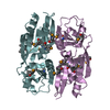
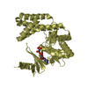
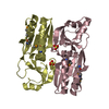
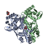
Crystal Structure of wild type MarR (apo form) from E.coli
Components
Multiple antibiotic resistance protein marR
Keywords
TRANSCRIPTION / winged helix-turn-helix DNA binding domain
Function / homology
Function and homology information
cellular response to antibiotic / response to stress / response to heat / DNA-binding transcription factor activity / negative regulation of DNA-templated transcription / regulation of DNA-templated transcription / DNA binding Similarity search - Function
Protocol: SINGLE WAVELENGTH / Monochromatic (M) / Laue (L): M / Scattering type: x-ray
Radiation wavelength
Wavelength: 1 Å / Relative weight: 1
Reflection
Resolution: 2.6→56.068 Å / Num. all: 8461 / Num. obs: 8461 / % possible obs: 99.9 % / Redundancy: 7.9 % / Rsym value: 0.07 / Net I/σ(I): 16.2
Reflection shell
Diffraction-ID: 1
Resolution (Å)
Redundancy (%)
Rmerge(I) all
Rmerge(I) obs
Mean I/σ(I) obs
Num. measured all
Num. unique all
Rpim(I) all
Rrim(I) all
Rsym value
Net I/σ(I) obs
% possible all
2.6-2.74
8.1
0.688
0.644
1.2
9690
1193
0.239
0.688
0.644
2.7
100
2.74-2.91
8
0.467
0.437
1.8
9078
1128
0.163
0.467
0.437
4.1
100
2.91-3.11
8
0.262
0.244
3.2
8608
1080
0.092
0.262
0.244
7.1
100
3.11-3.36
7.9
0.136
0.127
6.1
7888
998
0.048
0.136
0.127
12.8
100
3.36-3.68
7.8
0.088
0.083
8.9
7339
942
0.031
0.088
0.083
18.7
100
3.68-4.11
7.8
0.062
0.058
12.4
6561
843
0.022
0.062
0.058
24.2
100
4.11-4.75
7.9
0.05
0.047
11.5
6072
765
0.018
0.05
0.047
31.4
100
4.75-5.81
7.9
0.084
0.079
7.6
5210
662
0.029
0.084
0.079
29.4
100
5.81-8.22
7.6
0.035
0.032
18.7
4007
529
0.012
0.035
0.032
29.6
100
8.22-35.828
6.6
0.023
0.021
27.7
2115
321
0.009
0.023
0.021
37.4
97.3
-
Phasing
Phasing
Method: molecular replacement
Phasing MR
Model details: Phaser MODE: MR_AUTO
Highest resolution
Lowest resolution
Rotation
3 Å
29.08 Å
Translation
3 Å
29.08 Å
-
Processing
Software
Name
Version
Classification
NB
MOSFLM
datareduction
SCALA
3.3.16
datascaling
PHASER
2.3.0
phasing
REFMAC
refinement
PDB_EXTRACT
3.1
dataextraction
Refinement
Method to determine structure: MOLECULAR REPLACEMENT / Resolution: 2.6→40 Å / Cor.coef. Fo:Fc: 0.938 / Cor.coef. Fo:Fc free: 0.908 / WRfactor Rfree: 0.2902 / WRfactor Rwork: 0.2413 / Occupancy max: 1 / Occupancy min: 1 / FOM work R set: 0.7514 / SU B: 39.26 / SU ML: 0.358 / SU R Cruickshank DPI: 0.3463 / SU Rfree: 0.4126 / Cross valid method: THROUGHOUT / σ(F): 0 / ESU R Free: 0.413 / Stereochemistry target values: MAXIMUM LIKELIHOOD Details: HYDROGENS HAVE BEEN USED IF PRESENT IN THE INPUT U VALUES
Rfactor
Num. reflection
% reflection
Selection details
Rfree
0.2902
823
9.8 %
RANDOM
Rwork
0.2413
-
-
-
obs
0.246
8400
99.67 %
-
Solvent computation
Ion probe radii: 0.8 Å / Shrinkage radii: 0.8 Å / VDW probe radii: 1.2 Å / Solvent model: BABINET MODEL WITH MASK
In the structure databanks used in Yorodumi, some data are registered as the other names, "COVID-19 virus" and "2019-nCoV". Here are the details of the virus and the list of structure data.
Jan 31, 2019. EMDB accession codes are about to change! (news from PDBe EMDB page)
EMDB accession codes are about to change! (news from PDBe EMDB page)
The allocation of 4 digits for EMDB accession codes will soon come to an end. Whilst these codes will remain in use, new EMDB accession codes will include an additional digit and will expand incrementally as the available range of codes is exhausted. The current 4-digit format prefixed with “EMD-” (i.e. EMD-XXXX) will advance to a 5-digit format (i.e. EMD-XXXXX), and so on. It is currently estimated that the 4-digit codes will be depleted around Spring 2019, at which point the 5-digit format will come into force.
The EM Navigator/Yorodumi systems omit the EMD- prefix.
Related info.:Q: What is EMD? / ID/Accession-code notation in Yorodumi/EM Navigator
Yorodumi is a browser for structure data from EMDB, PDB, SASBDB, etc.
This page is also the successor to EM Navigator detail page, and also detail information page/front-end page for Omokage search.
The word "yorodu" (or yorozu) is an old Japanese word meaning "ten thousand". "mi" (miru) is to see.
Related info.:EMDB / PDB / SASBDB / Comparison of 3 databanks / Yorodumi Search / Aug 31, 2016. New EM Navigator & Yorodumi / Yorodumi Papers / Jmol/JSmol / Function and homology information / Changes in new EM Navigator and Yorodumi
 Movie
Movie Controller


 Open data
 Basic information
 Components
 Keywords
 Function and homology information
 X-RAY DIFFRACTION /
  Authors
 Citation
 Structure visualization
 Downloads & links
 Other downloads


 PDBj
PDBj Assembly
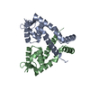
 Sample preparation
 / Beamline: 3W1A / Wavelength: 1 Å
 Processing