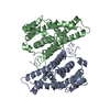
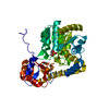
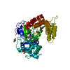
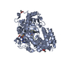
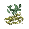
D: Bifunctional coenzyme PQQ synthesis protein C/D B: Bifunctional coenzyme PQQ synthesis protein C/D
defined by author&software
Evidence: homology, PqqC is a physiological monomer in most species (few are naturally fused as Methylobacterium extorquens PqqCD is). This truncation from the CD natural fusion is monomeric like most independent PqqCs.
Method: X-RAY DIFFRACTION / Number of used crystals: 1
-
Sample preparation
Crystal
Density Matthews: 2.9 Å3/Da / Density % sol: 57.52 %
Crystal grow
Temperature: 292 K / Method: vapor diffusion, hanging drop Details: 500 microL well volumes. Protein solution: 8.0 mg/mL protein, 50 mM Tris, pH 7.9, 100 mM sodium chloride, and 1 mM TCEP. Well solution: 100 mM HEPES, pH 6.7, 19% w/v PEG-4000, 10% ...Details: 500 microL well volumes. Protein solution: 8.0 mg/mL protein, 50 mM Tris, pH 7.9, 100 mM sodium chloride, and 1 mM TCEP. Well solution: 100 mM HEPES, pH 6.7, 19% w/v PEG-4000, 10% isopropanol. Water used in the well solutions contained 0.55 mM sodium azide. Hanging drops were 1 microL protein solution and 1 microL well solution
Resolution: 2.85→243.49 Å / Cor.coef. Fo:Fc: 0.914 / Cor.coef. Fo:Fc free: 0.854 / SU ML: 0.371 / Cross valid method: THROUGHOUT / ESU R: 0.995 / ESU R Free: 0.447 / Details: HYDROGENS HAVE BEEN ADDED IN THE RIDING POSITIONS
Rfactor
Num. reflection
% reflection
Selection details
Rfree
0.32707
1593
5.1 %
RANDOM
Rwork
0.25314
-
-
-
obs
0.2569
29899
98.38 %
-
Solvent computation
Ion probe radii: 0.8 Å / Shrinkage radii: 0.8 Å / VDW probe radii: 1.2 Å
Displacement parameters
Baniso -1
Baniso -2
Baniso -3
1-
1.09 Å2
0 Å2
0 Å2
2-
-
1.09 Å2
0 Å2
3-
-
-
-2.17 Å2
Refinement step
Cycle: 1 / Resolution: 2.85→243.49 Å
Protein
Nucleic acid
Ligand
Solvent
Total
Num. atoms
6570
0
0
0
6570
Refine LS restraints
Refine-ID
Type
Dev ideal
Dev ideal target
Number
X-RAY DIFFRACTION
r_bond_refined_d
0.018
0.019
6723
X-RAY DIFFRACTION
r_bond_other_d
0.002
0.02
6220
X-RAY DIFFRACTION
r_angle_refined_deg
2.088
1.941
9094
X-RAY DIFFRACTION
r_angle_other_deg
1.241
3
14247
X-RAY DIFFRACTION
r_dihedral_angle_1_deg
8.119
5
796
X-RAY DIFFRACTION
r_dihedral_angle_2_deg
37.886
22.312
333
X-RAY DIFFRACTION
r_dihedral_angle_3_deg
21.46
15
1091
X-RAY DIFFRACTION
r_dihedral_angle_4_deg
18.939
15
64
X-RAY DIFFRACTION
r_chiral_restr
0.119
0.2
990
X-RAY DIFFRACTION
r_gen_planes_refined
0.009
0.02
7425
X-RAY DIFFRACTION
r_gen_planes_other
0.002
0.02
1547
X-RAY DIFFRACTION
r_mcbond_it
3.615
5.984
3241
X-RAY DIFFRACTION
r_mcbond_other
3.613
5.985
3240
X-RAY DIFFRACTION
r_mcangle_it
5.64
8.963
4018
X-RAY DIFFRACTION
r_mcangle_other
5.641
8.963
4019
X-RAY DIFFRACTION
r_scbond_it
3.444
6.247
3482
X-RAY DIFFRACTION
r_scbond_other
3.444
6.247
3483
X-RAY DIFFRACTION
r_scangle_it
X-RAY DIFFRACTION
r_scangle_other
5.496
9.271
5077
X-RAY DIFFRACTION
r_long_range_B_refined
8.509
70.333
7957
X-RAY DIFFRACTION
r_long_range_B_other
8.503
70.323
7956
LS refinement shell
Resolution: 2.852→2.926 Å / Total num. of bins used: 20
Rfactor
Num. reflection
% reflection
Rfree
0.338
97
-
Rwork
0.335
1817
-
obs
-
-
82.89 %
Refinement TLS params.
Method: refined / Refine-ID: X-RAY DIFFRACTION
ID
L11 (°2)
L12 (°2)
L13 (°2)
L22 (°2)
L23 (°2)
L33 (°2)
S11 (Å °)
S12 (Å °)
S13 (Å °)
S21 (Å °)
S22 (Å °)
S23 (Å °)
S31 (Å °)
S32 (Å °)
S33 (Å °)
T11 (Å2)
T12 (Å2)
T13 (Å2)
T22 (Å2)
T23 (Å2)
T33 (Å2)
Origin x (Å)
Origin y (Å)
Origin z (Å)
1
1.3582
0.4162
0.0468
0.7923
-0.1204
1.0609
0.1034
-0.21
0.1472
0.0444
-0.1863
-0.0577
-0.0542
0.0405
0.0829
0.2392
-0.0516
-0.0185
0.2319
0.0158
0.0389
-36.619
6.6068
-20.8505
2
1.0019
-0.7245
0.4535
1.2526
-0.5712
0.8654
-0.0962
0.2478
0.0349
0.0614
-0.0318
-0.1243
0.057
0.1144
0.1281
0.1586
0.0251
0.0281
0.2485
-0.0196
0.0306
-35.1863
-11.788
15.7925
3
1.5991
0.7689
0.2293
1.123
-0.2252
0.4698
-0.0768
0.0576
0.2175
-0.1562
-0.0521
0.1188
-0.1399
0.0391
0.1289
0.2771
0.0067
-0.0557
0.1455
0.0185
0.0709
-52.216
16.1799
-41.1781
4
1.0038
0.6707
0.3419
1.091
0.5846
0.3179
0.2573
-0.4366
-0.04
0.14
-0.3138
0.0775
0.0853
-0.1793
0.0565
0.2727
-0.1617
-0.0105
0.3365
0.0195
0.0226
-62.8479
-17.6738
-16.6281
Refinement TLS group
ID
Refine-ID
Refine TLS-ID
Auth asym-ID
Auth seq-ID
1
X-RAY DIFFRACTION
1
D
15 - 251
2
X-RAY DIFFRACTION
2
A
16 - 251
3
X-RAY DIFFRACTION
3
B
17 - 248
4
X-RAY DIFFRACTION
4
C
19 - 244
+
About Yorodumi
-
News
-
Feb 9, 2022. New format data for meta-information of EMDB entries
New format data for meta-information of EMDB entries
Version 3 of the EMDB header file is now the official format.
The previous official version 1.9 will be removed from the archive.
In the structure databanks used in Yorodumi, some data are registered as the other names, "COVID-19 virus" and "2019-nCoV". Here are the details of the virus and the list of structure data.
Jan 31, 2019. EMDB accession codes are about to change! (news from PDBe EMDB page)
EMDB accession codes are about to change! (news from PDBe EMDB page)
The allocation of 4 digits for EMDB accession codes will soon come to an end. Whilst these codes will remain in use, new EMDB accession codes will include an additional digit and will expand incrementally as the available range of codes is exhausted. The current 4-digit format prefixed with “EMD-” (i.e. EMD-XXXX) will advance to a 5-digit format (i.e. EMD-XXXXX), and so on. It is currently estimated that the 4-digit codes will be depleted around Spring 2019, at which point the 5-digit format will come into force.
The EM Navigator/Yorodumi systems omit the EMD- prefix.
Related info.:Q: What is EMD? / ID/Accession-code notation in Yorodumi/EM Navigator
Yorodumi is a browser for structure data from EMDB, PDB, SASBDB, etc.
This page is also the successor to EM Navigator detail page, and also detail information page/front-end page for Omokage search.
The word "yorodu" (or yorozu) is an old Japanese word meaning "ten thousand". "mi" (miru) is to see.
Related info.:EMDB / PDB / SASBDB / Comparison of 3 databanks / Yorodumi Search / Aug 31, 2016. New EM Navigator & Yorodumi / Yorodumi Papers / Jmol/JSmol / Function and homology information / Changes in new EM Navigator and Yorodumi
 Movie
Movie Controller

 Yorodumi
 Open data
 Basic information
 Components
 Keywords
 Function and homology information
 Methylobacterium extorquens (bacteria)
 X-RAY DIFFRACTION /
  Authors
 United States, 2items
  Citation
 Structure visualization
 Downloads & links
 Other downloads


 PDBj
PDBj Assembly
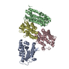

 Sample preparation
 Processing