 Movie
Movie Controller

[English] 日本語
 Yorodumi
- PDB-5vrc: Crystal structure for Methylobacterium extorquens PqqC (truncatio... -
+ Open data

- Basic information
| Entry | Database: PDB / ID: 5vrc | |||||||||
|---|---|---|---|---|---|---|---|---|---|---|
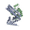
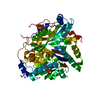
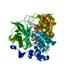
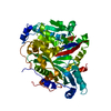
| Title | Crystal structure for Methylobacterium extorquens PqqC (truncation of natural CD fusion) | |||||||||
 Components Components | Bifunctional coenzyme PQQ synthesis protein C/D | |||||||||
 Keywords Keywords | OXIDOREDUCTASE / PQQ / oxidase / alpha-helical bundle | |||||||||
| Function / homology |  Function and homology information Function and homology informationpyrroloquinoline-quinone synthase / pyrroloquinoline-quinone synthase activity / pyrroloquinoline quinone biosynthetic process / quinone binding Similarity search - Function | |||||||||
| Biological species |  Methylobacterium extorquens (bacteria) Methylobacterium extorquens (bacteria) | |||||||||
| Method |  X-RAY DIFFRACTION / SYNCHROTRON / MOLECULAR REPLACEMENT / Resolution: 2 Å X-RAY DIFFRACTION / SYNCHROTRON / MOLECULAR REPLACEMENT / Resolution: 2 Å | |||||||||
 Authors Authors | Evans III, R.L. / Wilmot, C.M. / Esler, M.A. | |||||||||
| Funding support |  United States, 2items United States, 2items
| |||||||||
 Citation Citation | Journal: Not published Title: Crystal structures for Methylobacterium extorquens PqqC from the CD natural fusion and the C truncation Authors: Evans III, R.L. / Esler, M.A. / Latham, J.A. / Klinman, J.P. / Wilmot, C.M. | |||||||||
| History |
|
- Structure visualization
| Structure viewer | Molecule: MolmilJmol/JSmol |
|---|
- Downloads & links
-Download
| PDBx/mmCIF format | 5vrc.cif.gz | 357 KB | Display | PDBx/mmCIF format |
|---|---|---|---|---|
| PDB format | pdb5vrc.ent.gz | 288.3 KB | Display | PDB format |
| PDBx/mmJSON format | 5vrc.json.gz | Tree view | PDBx/mmJSON format | |
| Others |  Other downloads Other downloads |
-Validation report
| Arichive directory | https://data.pdbj.org/pub/pdb/validation_reports/vr/5vrcftp://data.pdbj.org/pub/pdb/validation_reports/vr/5vrc | HTTPS FTP |
|---|
-Related structure data
| Related structure data |  5vrdC  1otvS C: citing same article ( S: Starting model for refinement |
|---|---|
| Similar structure data |


-Links
 PDBj
PDBj- Assembly
| Deposited unit | 
| ||||||||
|---|---|---|---|---|---|---|---|---|---|
| 1 | 
| ||||||||
| 2 | 
| ||||||||
| Unit cell |
|
-Components
| #1: Protein | Mass: 31843.119 Da / Num. of mol.: 4 / Fragment: C domain residues 1-260 Source method: isolated from a genetically manipulated source Source: (gene. exp.) Methylobacterium extorquens (strain ATCC 14718 / DSM 1338 / JCM 2805 / NCIMB 9133 / AM1) (bacteria)Strain: ATCC 14718 / DSM 1338 / JCM 2805 / NCIMB 9133 / AM1 / Gene: pqqCD, MexAM1_META1p1749 / Production host: References: UniProt: Q49150, pyrroloquinoline-quinone synthase #2: Water | ChemComp-HOH / |  Mass: 18.015 Da / Num. of mol.: 84 / Source method: isolated from a natural source / Formula: H2O Mass: 18.015 Da / Num. of mol.: 84 / Source method: isolated from a natural source / Formula: H2O |
|---|
-Experimental details
-Experiment
| Experiment | Method: X-RAY DIFFRACTION / Number of used crystals: 1 |
|---|
- Sample preparation
| Crystal | Density Matthews: 2.04 Å3/Da / Density % sol: 39.61 % |
|---|---|
| Crystal grow | Temperature: 292 K / Method: vapor diffusion, hanging drop Details: 500 microL well volumes. Drops suspended on siliconized glass. Protein solution: 8.0 mg/mL protein in buffer (50 mM Tris, pH 8.0, 200mM sodium chloride). Well solution: 100 mM HEPES, pH 8. ...Details: 500 microL well volumes. Drops suspended on siliconized glass. Protein solution: 8.0 mg/mL protein in buffer (50 mM Tris, pH 8.0, 200mM sodium chloride). Well solution: 100 mM HEPES, pH 8.07, 200 mM sodium chloride, and 23.75% w/v PEG-3350. All water used in well solution buffers was 0.55 mM sodium azide for fungal growth suppression. Protein:well solution was 1:1 (1 microL to 1 microL) |
-Data collection
| Diffraction | Mean temperature: 100 K |
|---|---|
| Diffraction source | Source: SYNCHROTRON / Site: APS / Beamline: 23-ID-D / Wavelength: 1.0332 Å |
| Detector | Type: DECTRIS PILATUS3 6M / Detector: PIXEL / Date: Jul 18, 2015 |
| Radiation | Protocol: SINGLE WAVELENGTH / Monochromatic (M) / Laue (L): M / Scattering type: x-ray |
| Radiation wavelength | Wavelength: 1.0332 Å / Relative weight: 1 |
| Reflection | Resolution: 2→29.51 Å / Num. obs: 69169 / % possible obs: 97.8 % / Redundancy: 6.8 % / Rmerge(I) obs: 0.077 / Net I/σ(I): 14.4 |
- Processing
| Software |
| ||||||||||||||||||||||||||||||||||||||||||||||||||||||||||||||||||||||||||||||||||||||||||||||||||||||||||||||||||||||||||||||||||||||||||||||||||||||||||||||||||||||||||||||||||||||
|---|---|---|---|---|---|---|---|---|---|---|---|---|---|---|---|---|---|---|---|---|---|---|---|---|---|---|---|---|---|---|---|---|---|---|---|---|---|---|---|---|---|---|---|---|---|---|---|---|---|---|---|---|---|---|---|---|---|---|---|---|---|---|---|---|---|---|---|---|---|---|---|---|---|---|---|---|---|---|---|---|---|---|---|---|---|---|---|---|---|---|---|---|---|---|---|---|---|---|---|---|---|---|---|---|---|---|---|---|---|---|---|---|---|---|---|---|---|---|---|---|---|---|---|---|---|---|---|---|---|---|---|---|---|---|---|---|---|---|---|---|---|---|---|---|---|---|---|---|---|---|---|---|---|---|---|---|---|---|---|---|---|---|---|---|---|---|---|---|---|---|---|---|---|---|---|---|---|---|---|---|---|---|---|
| Refinement | Method to determine structure: MOLECULAR REPLACEMENT Starting model: 1otv Resolution: 2→29.51 Å / Cor.coef. Fo:Fc: 0.948 / Cor.coef. Fo:Fc free: 0.927 / SU B: 9.516 / SU ML: 0.122 / Cross valid method: THROUGHOUT / ESU R: 0.176 / ESU R Free: 0.159 / Stereochemistry target values: MAXIMUM LIKELIHOOD / Details: HYDROGENS HAVE BEEN ADDED IN THE RIDING POSITIONS
| ||||||||||||||||||||||||||||||||||||||||||||||||||||||||||||||||||||||||||||||||||||||||||||||||||||||||||||||||||||||||||||||||||||||||||||||||||||||||||||||||||||||||||||||||||||||
| Solvent computation | Ion probe radii: 0.8 Å / Shrinkage radii: 0.8 Å / VDW probe radii: 1.2 Å / Solvent model: MASK | ||||||||||||||||||||||||||||||||||||||||||||||||||||||||||||||||||||||||||||||||||||||||||||||||||||||||||||||||||||||||||||||||||||||||||||||||||||||||||||||||||||||||||||||||||||||
| Displacement parameters | Biso mean: 38.9 Å2
| ||||||||||||||||||||||||||||||||||||||||||||||||||||||||||||||||||||||||||||||||||||||||||||||||||||||||||||||||||||||||||||||||||||||||||||||||||||||||||||||||||||||||||||||||||||||
| Refinement step | Cycle: 1 / Resolution: 2→29.51 Å
| ||||||||||||||||||||||||||||||||||||||||||||||||||||||||||||||||||||||||||||||||||||||||||||||||||||||||||||||||||||||||||||||||||||||||||||||||||||||||||||||||||||||||||||||||||||||
| Refine LS restraints |
|