 Movie
Movie Controller

[English] 日本語
 Yorodumi
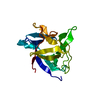
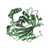
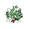
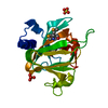
- PDB-5h0u: Crystal structure of the catalytic domain of membrane type 1 matr... -
+ Open data

- Basic information
| Entry | Database: PDB / ID: 5h0u | ||||||
|---|---|---|---|---|---|---|---|
| Title | Crystal structure of the catalytic domain of membrane type 1 matrix metalloproteinase | ||||||
 Components Components |
| ||||||
 Keywords Keywords | HYDROLASE / catalytic domain membrane type 1 matrix metalloproteinase MT1-MMP | ||||||
| Function / homology |  Function and homology information Function and homology informationmembrane-type matrix metalloproteinase-1 / negative regulation of GDF15-GFRAL signaling pathway / positive regulation of macrophage migration / craniofacial suture morphogenesis / macropinosome / response to odorant / chondrocyte proliferation / head development / astrocyte cell migration / TGFBR3 PTM regulation ...membrane-type matrix metalloproteinase-1 / negative regulation of GDF15-GFRAL signaling pathway / positive regulation of macrophage migration / craniofacial suture morphogenesis / macropinosome / response to odorant / chondrocyte proliferation / head development / astrocyte cell migration / TGFBR3 PTM regulation / tissue remodeling / negative regulation of focal adhesion assembly / positive regulation of protein processing / endochondral ossification / embryonic cranial skeleton morphogenesis / intermediate filament cytoskeleton / endothelial cell proliferation / zymogen activation / positive regulation of B cell differentiation / branching morphogenesis of an epithelial tube / positive regulation of myotube differentiation / Activation of Matrix Metalloproteinases / metalloaminopeptidase activity / endodermal cell differentiation / Collagen degradation / collagen catabolic process / extracellular matrix disassembly / negative regulation of Notch signaling pathway / response to mechanical stimulus / regulation of protein localization to plasma membrane / ovarian follicle development / Degradation of the extracellular matrix / extracellular matrix organization / extracellular matrix / lung development / skeletal system development / cell motility / protein catabolic process / metalloendopeptidase activity / protein processing / Golgi lumen / response to estrogen / male gonad development / integrin binding / melanosome / positive regulation of cell growth / response to oxidative stress / cytoplasmic vesicle / High laminar flow shear stress activates signaling by PIEZO1 and PECAM1:CDH5:KDR in endothelial cells / angiogenesis / endopeptidase activity / response to hypoxia / positive regulation of cell migration / serine-type endopeptidase activity / focal adhesion / proteolysis / extracellular space / zinc ion binding / nucleus / plasma membrane / cytosol Similarity search - Function | ||||||
| Biological species |  Homo sapiens (human) Homo sapiens (human)unidentified (others) | ||||||
| Method |  X-RAY DIFFRACTION / SYNCHROTRON / MOLECULAR REPLACEMENT / Resolution: 2.239 Å X-RAY DIFFRACTION / SYNCHROTRON / MOLECULAR REPLACEMENT / Resolution: 2.239 Å | ||||||
 Authors Authors | Ogata, H. / Decaneto, E. / Lubitz, W. | ||||||
 Citation Citation | Journal: Phys Chem Chem Phys / Year: 2017 Title: Solvent water interactions within the active site of the membrane type I matrix metalloproteinase. Authors: Decaneto, E. / Vasilevskaya, T. / Kutin, Y. / Ogata, H. / Grossman, M. / Sagi, I. / Havenith, M. / Lubitz, W. / Thiel, W. / Cox, N. | ||||||
| History |
|
- Structure visualization
| Structure viewer | Molecule: MolmilJmol/JSmol |
|---|
- Downloads & links
-Download
| PDBx/mmCIF format | 5h0u.cif.gz | 55.6 KB | Display | PDBx/mmCIF format |
|---|---|---|---|---|
| PDB format | pdb5h0u.ent.gz | 38.4 KB | Display | PDB format |
| PDBx/mmJSON format | 5h0u.json.gz | Tree view | PDBx/mmJSON format | |
| Others |  Other downloads Other downloads |
-Validation report
| Summary document | 5h0u_validation.pdf.gz | 457.1 KB | Display | wwPDB validaton report |
|---|---|---|---|---|
| Full document | 5h0u_full_validation.pdf.gz | 459.2 KB | Display | |
| Data in XML | 5h0u_validation.xml.gz | 10.9 KB | Display | |
| Data in CIF | 5h0u_validation.cif.gz | 13.9 KB | Display | |
| Arichive directory | https://data.pdbj.org/pub/pdb/validation_reports/h0/5h0uftp://data.pdbj.org/pub/pdb/validation_reports/h0/5h0u | HTTPS FTP |
-Related structure data
| Related structure data |  1bqqS S: Starting model for refinement |
|---|---|
| Similar structure data |


-Links
 PDBj
PDBj


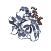
- Assembly
| Deposited unit | 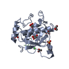
| ||||||||
|---|---|---|---|---|---|---|---|---|---|
| 1 |
| ||||||||
| Unit cell |
|
-Components
-Protein / Protein/peptide , 2 types, 2 molecules AB
| #1: Protein | Mass: 19338.320 Da / Num. of mol.: 1 Source method: isolated from a genetically manipulated source Source: (gene. exp.) Homo sapiens (human) / Gene: MMP14 / Production host:  References: UniProt: P50281, membrane-type matrix metalloproteinase-1 |
|---|---|
| #2: Protein/peptide | Mass: 846.896 Da / Num. of mol.: 1 Source method: isolated from a genetically manipulated source Details: His-tag / Source: (gene. exp.) unidentified (others) / Production host: |
-Non-polymers , 5 types, 82 molecules 


| #3: Chemical |  Mass: 40.078 Da / Num. of mol.: 2 / Source method: obtained synthetically / Formula: Ca Mass: 40.078 Da / Num. of mol.: 2 / Source method: obtained synthetically / Formula: Ca#4: Chemical |  Mass: 65.409 Da / Num. of mol.: 3 / Source method: obtained synthetically / Formula: Zn Mass: 65.409 Da / Num. of mol.: 3 / Source method: obtained synthetically / Formula: Zn#5: Chemical | ChemComp-EPE / |  Mass: 238.305 Da / Num. of mol.: 1 / Source method: isolated from a natural source / Formula: C8H18N2O4S / Comment: pH buffer*YM Mass: 238.305 Da / Num. of mol.: 1 / Source method: isolated from a natural source / Formula: C8H18N2O4S / Comment: pH buffer*YM#6: Chemical | ChemComp-GOL /  Mass: 92.094 Da / Num. of mol.: 8 / Source method: obtained synthetically / Formula: C3H8O3 Mass: 92.094 Da / Num. of mol.: 8 / Source method: obtained synthetically / Formula: C3H8O3#7: Water | ChemComp-HOH / | Mass: 18.015 Da / Num. of mol.: 68 / Source method: isolated from a natural source / Formula: H2O |
|---|
-Experimental details
-Experiment
| Experiment | Method: X-RAY DIFFRACTION / Number of used crystals: 1 |
|---|
- Sample preparation
| Crystal | Density Matthews: 3.04 Å3/Da / Density % sol: 59.49 % |
|---|---|
| Crystal grow | Temperature: 277 K / Method: vapor diffusion, sitting drop / Details: Ammonium nitrate, PEG3350 |
-Data collection
| Diffraction | Mean temperature: 100 K |
|---|---|
| Diffraction source | Source: SYNCHROTRON / Site: BESSY  / Beamline: 14.2 / Wavelength: 0.91841 Å / Beamline: 14.2 / Wavelength: 0.91841 Å |
| Detector | Type: RAYONIX MX225HE / Detector: CCD / Date: Aug 2, 2013 |
| Radiation | Protocol: SINGLE WAVELENGTH / Monochromatic (M) / Laue (L): M / Scattering type: x-ray |
| Radiation wavelength | Wavelength: 0.91841 Å / Relative weight: 1 |
| Reflection | Resolution: 2.239→44.544 Å / Num. obs: 12491 / % possible obs: 99.8 % / Redundancy: 7.9 % / Rmerge(I) obs: 0.105 / Net I/σ(I): 16.8 |
| Reflection shell | Resolution: 2.239→2.37 Å / Rmerge(I) obs: 0.998 / Mean I/σ(I) obs: 2.3 / % possible all: 99.3 |
- Processing
| Software |
| |||||||||||||||||||||||||||||||||||
|---|---|---|---|---|---|---|---|---|---|---|---|---|---|---|---|---|---|---|---|---|---|---|---|---|---|---|---|---|---|---|---|---|---|---|---|---|
| Refinement | Method to determine structure: MOLECULAR REPLACEMENT Starting model: 1BQQ Resolution: 2.239→44.544 Å / SU ML: 0.28 / Cross valid method: FREE R-VALUE / σ(F): 1.35 / Phase error: 26.11
| |||||||||||||||||||||||||||||||||||
| Solvent computation | Shrinkage radii: 0.9 Å / VDW probe radii: 1.11 Å | |||||||||||||||||||||||||||||||||||
| Refinement step | Cycle: LAST / Resolution: 2.239→44.544 Å
| |||||||||||||||||||||||||||||||||||
| Refine LS restraints |
| |||||||||||||||||||||||||||||||||||
| LS refinement shell |
|
