 Movie
Movie Controller

[English] 日本語
 Yorodumi
- PDB-5fuo: Extending the half-life of a Fab fragment through generation of a... -
+ Open data

- Basic information
| Entry | Database: PDB / ID: 5fuo | ||||||
|---|---|---|---|---|---|---|---|
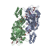
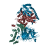
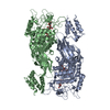
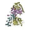
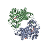
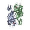
| Title | Extending the half-life of a Fab fragment through generation of a humanised anti-Human Serum Albumin (HSA) Fv domain: an investigation into the correlation between affinity and serum half-life | ||||||
 Components Components |
| ||||||
 Keywords Keywords | IMMUNE SYSTEM / ANTI-ALBUMIN / FAB FRAGMENT / SERUM HALF-LIFE / FCRN / HUMAN SERUM ALBUMIN | ||||||
| Function / homology |  Function and homology information Function and homology informationbilirubin transport / Ciprofloxacin ADME / exogenous protein binding / cellular response to calcium ion starvation / enterobactin binding / Heme biosynthesis / HDL remodeling / molecular carrier activity / negative regulation of mitochondrial depolarization / Prednisone ADME ...bilirubin transport / Ciprofloxacin ADME / exogenous protein binding / cellular response to calcium ion starvation / enterobactin binding / Heme biosynthesis / HDL remodeling / molecular carrier activity / negative regulation of mitochondrial depolarization / Prednisone ADME / Heme degradation / Aspirin ADME / antioxidant activity / Scavenging of heme from plasma / toxic substance binding / Recycling of bile acids and salts / platelet alpha granule lumen / fatty acid binding / cellular response to starvation / Post-translational protein phosphorylation / Cytoprotection by HMOX1 / response to nutrient levels / Regulation of Insulin-like Growth Factor (IGF) transport and uptake by Insulin-like Growth Factor Binding Proteins (IGFBPs) / Platelet degranulation / pyridoxal phosphate binding / protein-folding chaperone binding / blood microparticle / endoplasmic reticulum lumen / copper ion binding / Golgi apparatus / endoplasmic reticulum / protein-containing complex / : / DNA binding / extracellular exosome / extracellular region / identical protein binding / nucleus Similarity search - Function | ||||||
| Biological species |  HOMO SAPIENS (human) HOMO SAPIENS (human) | ||||||
| Method |  X-RAY DIFFRACTION / SYNCHROTRON / MOLECULAR REPLACEMENT / Resolution: 3.6 Å X-RAY DIFFRACTION / SYNCHROTRON / MOLECULAR REPLACEMENT / Resolution: 3.6 Å | ||||||
 Authors Authors | Adams, R. / Ceska, T. | ||||||
 Citation Citation | Journal: Mabs / Year: 2016 Title: Extending the Half-Life of a Fab Fragment Through Generation of a Humanized Anti-Human Serum Albumin Fv Domain: An Investigation Into the Correlation between Affinity and Serum Half-Life. Authors: Adams, R. / Griffin, L. / Compson, J.E. / Jairaj, M. / Baker, T. / Ceska, T. / West, S. / Zaccheo, O. / Dave, E. / Lawson, A.D.G. / Humphreys, D.P. / Heywood, S. | ||||||
| History |
|
- Structure visualization
| Structure viewer | Molecule: MolmilJmol/JSmol |
|---|
- Downloads & links
-Download
| PDBx/mmCIF format | 5fuo.cif.gz | 205.1 KB | Display | PDBx/mmCIF format |
|---|---|---|---|---|
| PDB format | pdb5fuo.ent.gz | 163.5 KB | Display | PDB format |
| PDBx/mmJSON format | 5fuo.json.gz | Tree view | PDBx/mmJSON format | |
| Others |  Other downloads Other downloads |
-Validation report
| Arichive directory | https://data.pdbj.org/pub/pdb/validation_reports/fu/5fuoftp://data.pdbj.org/pub/pdb/validation_reports/fu/5fuo | HTTPS FTP |
|---|
-Related structure data
| Related structure data |  5fuzC  4g03S C: citing same article ( S: Starting model for refinement |
|---|---|
| Similar structure data |


-Links
 PDBj
PDBj


- Assembly
| Deposited unit | 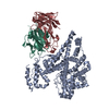
| ||||||||
|---|---|---|---|---|---|---|---|---|---|
| 1 |
| ||||||||
| Unit cell |
|
-Components
| #1: Protein | Mass: 66571.219 Da / Num. of mol.: 1 Source method: isolated from a genetically manipulated source Source: (gene. exp.) HOMO SAPIENS (human) / Organ: TESTIS / Cell line (production host): CHO / Production host:   CRICETULUS GRISEUS (Chinese hamster) / References: UniProt: P02768 CRICETULUS GRISEUS (Chinese hamster) / References: UniProt: P02768 |
|---|---|
| #2: Antibody | Mass: 24900.941 Da / Num. of mol.: 1 Source method: isolated from a genetically manipulated source Source: (gene. exp.) HOMO SAPIENS (human) / Organ: TESTIS / Cell line (production host): CHO / Production host: CRICETULUS GRISEUS (Chinese hamster) |
| #3: Antibody | Mass: 23419.875 Da / Num. of mol.: 1 Source method: isolated from a genetically manipulated source Source: (gene. exp.) HOMO SAPIENS (human) / Organ: TESTIS / Cell line (production host): CHO / Production host: CRICETULUS GRISEUS (Chinese hamster) |
| Has protein modification | Y |
| Sequence details | IN-HOUSE DISCOVERED |
-Experimental details
-Experiment
| Experiment | Method: X-RAY DIFFRACTION / Number of used crystals: 1 |
|---|
- Sample preparation
| Crystal | Density Matthews: 4.72 Å3/Da / Density % sol: 73.94 % / Description: NONE |
|---|---|
| Crystal grow | pH: 1 Details: 0.1 M CITRIC ACID PH 4.4, 0.1 M DI-SODIUM HYDROGEN PHOSPHATE, 38% V/V ETHANOL, 5% V/V POLYETHYLENE GLYCOL 1000 |
-Data collection
| Diffraction | Mean temperature: 100 K |
|---|---|
| Diffraction source | Source: SYNCHROTRON / Site: Diamond  / Beamline: I02 / Wavelength: 0.9713 / Beamline: I02 / Wavelength: 0.9713 |
| Detector | Type: DECTRIS PILATUS / Detector: PIXEL / Date: Mar 23, 2012 |
| Radiation | Protocol: SINGLE WAVELENGTH / Monochromatic (M) / Laue (L): M / Scattering type: x-ray |
| Radiation wavelength | Wavelength: 0.9713 Å / Relative weight: 1 |
| Reflection | Resolution: 3.58→30 Å / Num. obs: 46115 / % possible obs: 93.9 % / Observed criterion σ(I): 1.6 / Redundancy: 2.4 % / Rmerge(I) obs: 0.01 / Net I/σ(I): 7.57 |
| Reflection shell | Resolution: 3.58→3.79 Å / Redundancy: 1.9 % / Rmerge(I) obs: 0.04 / Mean I/σ(I) obs: 1.64 / % possible all: 86.1 |
- Processing
| Software |
| ||||||||||||||||||||||||||||||||||||||||||||||||||||||||||||||||||||||||||||||||
|---|---|---|---|---|---|---|---|---|---|---|---|---|---|---|---|---|---|---|---|---|---|---|---|---|---|---|---|---|---|---|---|---|---|---|---|---|---|---|---|---|---|---|---|---|---|---|---|---|---|---|---|---|---|---|---|---|---|---|---|---|---|---|---|---|---|---|---|---|---|---|---|---|---|---|---|---|---|---|---|---|---|
| Refinement | Method to determine structure: MOLECULAR REPLACEMENT Starting model: PDB ENTRY 4G03 AND FAB WWPDB SUBMISSION CODE 62161 Resolution: 3.6→30 Å / Rfactor Rfree error: 0.005 / Data cutoff high absF: 10000 / Data cutoff low absF: 0 / Isotropic thermal model: RESTRAINED / Cross valid method: THROUGHOUT / σ(F): 0 Details: CHAIN A RESIDUES 1-4, 583-585 AND CHAIN H RESIDUES 136-142, 224-233 ARE TOO DISORDERED TO MODEL
| ||||||||||||||||||||||||||||||||||||||||||||||||||||||||||||||||||||||||||||||||
| Solvent computation | Bsol: 64.83 Å2 / ksol: 0.3 e/Å3 | ||||||||||||||||||||||||||||||||||||||||||||||||||||||||||||||||||||||||||||||||
| Displacement parameters |
| ||||||||||||||||||||||||||||||||||||||||||||||||||||||||||||||||||||||||||||||||
| Refinement step | Cycle: LAST / Resolution: 3.6→30 Å
| ||||||||||||||||||||||||||||||||||||||||||||||||||||||||||||||||||||||||||||||||
| Refine LS restraints |
| ||||||||||||||||||||||||||||||||||||||||||||||||||||||||||||||||||||||||||||||||
| LS refinement shell | Resolution: 3.6→3.73 Å / Rfactor Rfree error: 0.007 / Total num. of bins used: 10
| ||||||||||||||||||||||||||||||||||||||||||||||||||||||||||||||||||||||||||||||||
| Xplor file |
|
