 Movie
Movie Controller

[English] 日本語
 Yorodumi
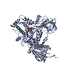
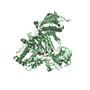
- PDB-4jaw: Crystal Structure of Lacto-N-Biosidase from Bifidobacterium bifid... -
+ Open data

- Basic information
| Entry | Database: PDB / ID: 4jaw | ||||||
|---|---|---|---|---|---|---|---|
| Title | Crystal Structure of Lacto-N-Biosidase from Bifidobacterium bifidum complexed with LNB-thiazoline | ||||||
 Components Components | Lacto-N-biosidase | ||||||
 Keywords Keywords | HYDROLASE / alpha/beta-domain / Tim Barrel / beta-Trefoil / Membrane-anchored extracellular | ||||||
| Function / homology |  Function and homology information Function and homology informationlacto-N-biosidase activity / lacto-N-biosidase / beta-N-acetylhexosaminidase activity / carbohydrate metabolic process / plasma membrane Similarity search - Function | ||||||
| Biological species |  Bifidobacterium bifidum (bacteria) Bifidobacterium bifidum (bacteria) | ||||||
| Method |  X-RAY DIFFRACTION / SYNCHROTRON / FOURIER SYNTHESIS / Resolution: 1.8 Å X-RAY DIFFRACTION / SYNCHROTRON / FOURIER SYNTHESIS / Resolution: 1.8 Å | ||||||
 Authors Authors | Ito, T. / Katayama, T. / Stubbs, K.A. / Fushinobu, S. | ||||||
 Citation Citation | Journal: J.Biol.Chem. / Year: 2013 Title: Crystal structures of a glycoside hydrolase family 20 lacto-N-biosidase from Bifidobacterium bifidum Authors: Ito, T. / Katayama, T. / Hattie, M. / Sakurama, H. / Wada, J. / Suzuki, R. / Ashida, H. / Wakagi, T. / Yamamoto, K. / Stubbs, K.A. / Fushinobu, S. #1: Journal: Appl.Environ.Microbiol. / Year: 2008 Title: Bifidobacterium bifidum lacto-N-biosidase, a critical enzyme for the degradation of human milk oligosaccharides with a type 1 structure. Authors: Wada, J. / Ando, T. / Kiyohara, M. / Ashida, H. / Kitaoka, M. / Yamaguchi, M. / Kumagai, H. / Katayama, T. / Yamamoto, K. | ||||||
| History |
|
- Structure visualization
| Structure viewer | Molecule: MolmilJmol/JSmol |
|---|
- Downloads & links
-Download
| PDBx/mmCIF format | 4jaw.cif.gz | 263 KB | Display | PDBx/mmCIF format |
|---|---|---|---|---|
| PDB format | pdb4jaw.ent.gz | 209.8 KB | Display | PDB format |
| PDBx/mmJSON format | 4jaw.json.gz | Tree view | PDBx/mmJSON format | |
| Others |  Other downloads Other downloads |
-Validation report
| Arichive directory | https://data.pdbj.org/pub/pdb/validation_reports/ja/4jawftp://data.pdbj.org/pub/pdb/validation_reports/ja/4jaw | HTTPS FTP |
|---|
-Related structure data
| Related structure data |  4h04SC S: Starting model for refinement C: citing same article ( |
|---|---|
| Similar structure data |


-Links
 PDBj
PDBj


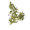
- Assembly
| Deposited unit | 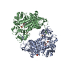
| |||||||||
|---|---|---|---|---|---|---|---|---|---|---|
| 1 | 
| |||||||||
| 2 | 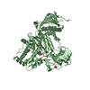
| |||||||||
| 3 |
| |||||||||
| Unit cell |
| |||||||||
| Components on special symmetry positions |
|
-Components
| #1: Protein | Mass: 71892.227 Da / Num. of mol.: 2 / Fragment: UNP Residue 41-663 Source method: isolated from a genetically manipulated source Source: (gene. exp.) Bifidobacterium bifidum (bacteria) / Strain: JCM1254 / Gene: lnbB / Plasmid: pET-28b / Production host: #2: Sugar |   Type: D-saccharide, beta linking / Mass: 180.156 Da / Num. of mol.: 2 Type: D-saccharide, beta linking / Mass: 180.156 Da / Num. of mol.: 2Source method: isolated from a genetically manipulated source Formula: C6H12O6 #3: Chemical |   Mass: 219.258 Da / Num. of mol.: 2 / Source method: obtained synthetically / Formula: C8H13NO4S Mass: 219.258 Da / Num. of mol.: 2 / Source method: obtained synthetically / Formula: C8H13NO4S#4: Chemical | ChemComp-SO4 /   Mass: 96.063 Da / Num. of mol.: 7 / Source method: obtained synthetically / Formula: SO4 Mass: 96.063 Da / Num. of mol.: 7 / Source method: obtained synthetically / Formula: SO4#5: Water | ChemComp-HOH / |  Mass: 18.015 Da / Num. of mol.: 403 / Source method: isolated from a natural source / Formula: H2O Mass: 18.015 Da / Num. of mol.: 403 / Source method: isolated from a natural source / Formula: H2OHas protein modification | Y | |
|---|
-Experimental details
-Experiment
| Experiment | Method: X-RAY DIFFRACTION / Number of used crystals: 1 |
|---|
- Sample preparation
| Crystal | Density Matthews: 2.79 Å3/Da / Density % sol: 55.95 % |
|---|---|
| Crystal grow | Temperature: 293 K / Method: vapor diffusion, sitting drop / pH: 5.6 Details: 0.2M potassium sodium tartrate tetrahydrate, 0.1M sodium citrate, 2.0M ammonium sulfate, pH 5.6, VAPOR DIFFUSION, SITTING DROP, temperature 293K |
-Data collection
| Diffraction | Mean temperature: 100 K |
|---|---|
| Diffraction source | Source: SYNCHROTRON / Site: Photon Factory  / Beamline: BL-17A / Wavelength: 1 Å / Beamline: BL-17A / Wavelength: 1 Å |
| Detector | Type: ADSC QUANTUM 4r / Detector: CCD / Date: Nov 25, 2012 |
| Radiation | Monochromator: Si(111), numerical link type double crystal monochromator Protocol: SINGLE WAVELENGTH / Monochromatic (M) / Laue (L): M / Scattering type: x-ray |
| Radiation wavelength | Wavelength: 1 Å / Relative weight: 1 |
| Reflection | Resolution: 1.8→50 Å / Num. obs: 150089 / % possible obs: 100 % / Observed criterion σ(I): 0 / Redundancy: 7.4 % / Biso Wilson estimate: 12.1 Å2 / Rsym value: 0.102 / Net I/σ(I): 23.97 |
| Reflection shell | Resolution: 1.8→1.83 Å / Redundancy: 7.3 % / Mean I/σ(I) obs: 4.85 / Num. unique all: 7436 / Rsym value: 0.485 / % possible all: 100 |
- Processing
| Software |
| |||||||||||||||||||||||||||||||||||||||||||||
|---|---|---|---|---|---|---|---|---|---|---|---|---|---|---|---|---|---|---|---|---|---|---|---|---|---|---|---|---|---|---|---|---|---|---|---|---|---|---|---|---|---|---|---|---|---|---|
| Refinement | Method to determine structure: FOURIER SYNTHESIS Starting model: PDB ENTRY 4H04 Resolution: 1.8→43.61 Å / Cor.coef. Fo:Fc: 0.941 / Cor.coef. Fo:Fc free: 0.925 / SU B: 1.879 / SU ML: 0.061 / Cross valid method: THROUGHOUT / ESU R: 0.107 / ESU R Free: 0.106 / Stereochemistry target values: MAXIMUM LIKELIHOOD / Details: HYDROGENS HAVE BEEN USED IF PRESENT IN THE INPUT
| |||||||||||||||||||||||||||||||||||||||||||||
| Solvent computation | Ion probe radii: 0.8 Å / Shrinkage radii: 0.8 Å / VDW probe radii: 1.2 Å / Solvent model: MASK | |||||||||||||||||||||||||||||||||||||||||||||
| Displacement parameters | Biso mean: 11.513 Å2
| |||||||||||||||||||||||||||||||||||||||||||||
| Refine analyze |
| |||||||||||||||||||||||||||||||||||||||||||||
| Refinement step | Cycle: LAST / Resolution: 1.8→43.61 Å
| |||||||||||||||||||||||||||||||||||||||||||||
| Refine LS restraints |
| |||||||||||||||||||||||||||||||||||||||||||||
| LS refinement shell | Resolution: 1.8→1.847 Å / Total num. of bins used: 20
|
