 Movie
Movie Controller

[English] 日本語
 Yorodumi
- PDB-5f4x: Fructose-1,6-bisphosphate aldolase K229M mutant from rabbit muscle -
+ Open data

- Basic information
| Entry | Database: PDB / ID: 5f4x | ||||||
|---|---|---|---|---|---|---|---|
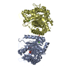
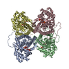
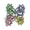
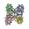
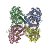
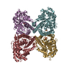
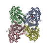
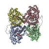
| Title | Fructose-1,6-bisphosphate aldolase K229M mutant from rabbit muscle | ||||||
 Components Components | Fructose-bisphosphate aldolase A | ||||||
 Keywords Keywords | LYASE / Aldolase / Class I / Lysine mutant | ||||||
| Function / homology |  Function and homology information Function and homology informationnegative regulation of Arp2/3 complex-mediated actin nucleation / fructose-bisphosphate aldolase / fructose-bisphosphate aldolase activity / M band / I band / glycolytic process / protein homotetramerization / positive regulation of cell migration Similarity search - Function | ||||||
| Biological species |  | ||||||
| Method |  X-RAY DIFFRACTION / SYNCHROTRON / MOLECULAR REPLACEMENT / Resolution: 1.84 Å X-RAY DIFFRACTION / SYNCHROTRON / MOLECULAR REPLACEMENT / Resolution: 1.84 Å | ||||||
 Authors Authors | LowKam, C. / Arthus-Cartier, G. | ||||||
 Citation Citation | Journal: To Be Published Title: DECYCLIZATION DETERMINES DIASTEREOISOMERIC SUBSTRATE SPECIFICITY OF MAMMALIAN CLASS I FRUCTOSE-1,6-BISPHOSPHATE ALDOLASE Authors: LowKam, C. / Arthus-Cartier, G. / Sygusch, J. | ||||||
| History |
|
- Structure visualization
| Structure viewer | Molecule: MolmilJmol/JSmol |
|---|
- Downloads & links
-Download
| PDBx/mmCIF format | 5f4x.cif.gz | 558.6 KB | Display | PDBx/mmCIF format |
|---|---|---|---|---|
| PDB format | pdb5f4x.ent.gz | 464.8 KB | Display | PDB format |
| PDBx/mmJSON format | 5f4x.json.gz | Tree view | PDBx/mmJSON format | |
| Others |  Other downloads Other downloads |
-Validation report
| Arichive directory | https://data.pdbj.org/pub/pdb/validation_reports/f4/5f4xftp://data.pdbj.org/pub/pdb/validation_reports/f4/5f4x | HTTPS FTP |
|---|
-Related structure data
| Related structure data | 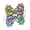 1zahS  5f2j S: Starting model for refinement |
|---|---|
| Similar structure data |


-Links
 PDBj
PDBj
- Assembly
| Deposited unit | 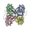
| ||||||||
|---|---|---|---|---|---|---|---|---|---|
| 1 |
| ||||||||
| Unit cell |
|
-Components
| #1: Protein | Mass: 39265.688 Da / Num. of mol.: 4 / Mutation: K229M Source method: isolated from a genetically manipulated source Source: (gene. exp.)  #2: Chemical |   Mass: 92.094 Da / Num. of mol.: 2 / Source method: obtained synthetically / Formula: C3H8O3 Mass: 92.094 Da / Num. of mol.: 2 / Source method: obtained synthetically / Formula: C3H8O3#3: Water | ChemComp-HOH / |  Mass: 18.015 Da / Num. of mol.: 1834 / Source method: isolated from a natural source / Formula: H2O Mass: 18.015 Da / Num. of mol.: 1834 / Source method: isolated from a natural source / Formula: H2O |
|---|
-Experimental details
-Experiment
| Experiment | Method: X-RAY DIFFRACTION / Number of used crystals: 1 |
|---|
- Sample preparation
| Crystal | Density Matthews: 2.29 Å3/Da / Density % sol: 46.33 % |
|---|---|
| Crystal grow | Temperature: 296 K / Method: vapor diffusion, hanging drop / pH: 7.5 Details: Sodium HEPES, PEG 4000, pH 7.5, VAPOR DIFFUSION, HANGING DROP, temperature 296K |
-Data collection
| Diffraction | Mean temperature: 100 K |
|---|---|
| Diffraction source | Source: SYNCHROTRON / Site: NSLS  / Beamline: X29A / Wavelength: 0.9795 Å / Beamline: X29A / Wavelength: 0.9795 Å |
| Detector | Type: ADSC QUANTUM 315 / Detector: CCD / Date: Jul 18, 2007 |
| Radiation | Protocol: SINGLE WAVELENGTH / Monochromatic (M) / Laue (L): M / Scattering type: x-ray |
| Radiation wavelength | Wavelength: 0.9795 Å / Relative weight: 1 |
| Reflection | Resolution: 1.84→35.201 Å / Num. obs: 122560 / % possible obs: 99.7 % / Redundancy: 3.6 % / Net I/σ(I): 25.13 |
| Reflection shell | Resolution: 1.84→1.91 Å / Redundancy: 3.1 % / Mean I/σ(I) obs: 3.43 / Num. unique all: 12223 / % possible all: 99.7 |
- Processing
| Software |
| |||||||||||||||||||||||||||||||||||||||||||||||||||||||||||||||||||||||||||||||||||||||||||||||||||||||||
|---|---|---|---|---|---|---|---|---|---|---|---|---|---|---|---|---|---|---|---|---|---|---|---|---|---|---|---|---|---|---|---|---|---|---|---|---|---|---|---|---|---|---|---|---|---|---|---|---|---|---|---|---|---|---|---|---|---|---|---|---|---|---|---|---|---|---|---|---|---|---|---|---|---|---|---|---|---|---|---|---|---|---|---|---|---|---|---|---|---|---|---|---|---|---|---|---|---|---|---|---|---|---|---|---|---|---|
| Refinement | Method to determine structure: MOLECULAR REPLACEMENT Starting model: 1ZAH Resolution: 1.84→35.201 Å / SU ML: 0.15 / Cross valid method: FREE R-VALUE / σ(F): 1.35 / Phase error: 16.83 / Stereochemistry target values: ML
| |||||||||||||||||||||||||||||||||||||||||||||||||||||||||||||||||||||||||||||||||||||||||||||||||||||||||
| Solvent computation | Shrinkage radii: 0.9 Å / VDW probe radii: 1.11 Å / Solvent model: FLAT BULK SOLVENT MODEL | |||||||||||||||||||||||||||||||||||||||||||||||||||||||||||||||||||||||||||||||||||||||||||||||||||||||||
| Refinement step | Cycle: LAST / Resolution: 1.84→35.201 Å
| |||||||||||||||||||||||||||||||||||||||||||||||||||||||||||||||||||||||||||||||||||||||||||||||||||||||||
| Refine LS restraints |
| |||||||||||||||||||||||||||||||||||||||||||||||||||||||||||||||||||||||||||||||||||||||||||||||||||||||||
| LS refinement shell |
|
