 Movie
Movie Controller

[English] 日本語
 Yorodumi
- PDB-1zal: Fructose-1,6-bisphosphate aldolase from rabbit muscle in complex ... -
+ Open data

- Basic information
| Entry | Database: PDB / ID: 1zal | ||||||
|---|---|---|---|---|---|---|---|
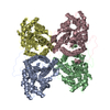
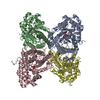
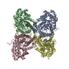
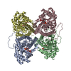
| Title | Fructose-1,6-bisphosphate aldolase from rabbit muscle in complex with partially disordered tagatose-1,6-bisphosphate, a weak competitive inhibitor | ||||||
 Components Components | Fructose-bisphosphate aldolase A | ||||||
 Keywords Keywords | LYASE / aldolase / weak competitive inhibitor / non covalent complex / tagatose-1 / 6-bisphosphate | ||||||
| Function / homology |  Function and homology information Function and homology informationnegative regulation of Arp2/3 complex-mediated actin nucleation / fructose-bisphosphate aldolase / fructose-bisphosphate aldolase activity / M band / I band / glycolytic process / protein homotetramerization / positive regulation of cell migration Similarity search - Function | ||||||
| Biological species |  | ||||||
| Method |  X-RAY DIFFRACTION / SYNCHROTRON / MOLECULAR REPLACEMENT / Resolution: 1.89 Å X-RAY DIFFRACTION / SYNCHROTRON / MOLECULAR REPLACEMENT / Resolution: 1.89 Å | ||||||
 Authors Authors | St-Jean, M. / Lafrance-Vanasse, J. / Liotard, B. / Sygusch, J. | ||||||
 Citation Citation | Journal: J.Biol.Chem. / Year: 2005 Title: High Resolution Reaction Intermediates of Rabbit Muscle Fructose-1,6-bisphosphate Aldolase: substrate cleavage and induced fit. Authors: St-Jean, M. / Lafrance-Vanasse, J. / Liotard, B. / Sygusch, J. | ||||||
| History |
| ||||||
| Remark 600 | HETEROGEN Electron density associated with tagatose-1,6-bisphosphate was not clearly defined and ...HETEROGEN Electron density associated with tagatose-1,6-bisphosphate was not clearly defined and only phosphate groups that were visible in the active sites of the enzyme were refined as PO4 ions. |
- Structure visualization
| Structure viewer | Molecule: MolmilJmol/JSmol |
|---|
- Downloads & links
-Download
| PDBx/mmCIF format | 1zal.cif.gz | 334.1 KB | Display | PDBx/mmCIF format |
|---|---|---|---|---|
| PDB format | pdb1zal.ent.gz | 266.9 KB | Display | PDB format |
| PDBx/mmJSON format | 1zal.json.gz | Tree view | PDBx/mmJSON format | |
| Others |  Other downloads Other downloads |
-Validation report
| Arichive directory | https://data.pdbj.org/pub/pdb/validation_reports/za/1zalftp://data.pdbj.org/pub/pdb/validation_reports/za/1zal | HTTPS FTP |
|---|
-Related structure data
| Related structure data | 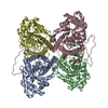 1zahC 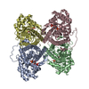 1zaiC  1zajC 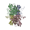 1adoS C: citing same article ( S: Starting model for refinement |
|---|---|
| Similar structure data |


-Links
 PDBj
PDBj
- Assembly
| Deposited unit | 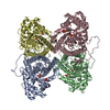
| ||||||||
|---|---|---|---|---|---|---|---|---|---|
| 1 |
| ||||||||
| Unit cell |
| ||||||||
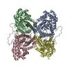
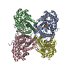
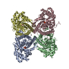
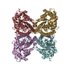
| Details | The biological assembly is the homotetramer found in the asymmetric unit |
-Components
| #1: Protein | Mass: 39263.672 Da / Num. of mol.: 4 Source method: isolated from a genetically manipulated source Source: (gene. exp.)  #2: Chemical | ChemComp-PO4 /   Mass: 94.971 Da / Num. of mol.: 8 / Source method: obtained synthetically / Formula: PO4 Mass: 94.971 Da / Num. of mol.: 8 / Source method: obtained synthetically / Formula: PO4#3: Water | ChemComp-HOH / |  Mass: 18.015 Da / Num. of mol.: 2215 / Source method: isolated from a natural source / Formula: H2O Mass: 18.015 Da / Num. of mol.: 2215 / Source method: isolated from a natural source / Formula: H2O |
|---|
-Experimental details
-Experiment
| Experiment | Method: X-RAY DIFFRACTION / Number of used crystals: 1 |
|---|
- Sample preparation
| Crystal | Density Matthews: 2.28 Å3/Da / Density % sol: 46.1 % |
|---|---|
| Crystal grow | Temperature: 296 K / Method: vapor diffusion, hanging drop / pH: 7.5 Details: sodium HEPES, PEG 4000, pH 7.5, VAPOR DIFFUSION, HANGING DROP, temperature 296K |
-Data collection
| Diffraction | Mean temperature: 100 K |
|---|---|
| Diffraction source | Source: SYNCHROTRON / Site: NSLS  / Beamline: X8C / Wavelength: 1 Å / Beamline: X8C / Wavelength: 1 Å |
| Detector | Type: ADSC QUANTUM 4 / Detector: CCD / Date: Feb 1, 2004 |
| Radiation | Protocol: SINGLE WAVELENGTH / Monochromatic (M) / Laue (L): M / Scattering type: x-ray |
| Radiation wavelength | Wavelength: 1 Å / Relative weight: 1 |
| Reflection | Resolution: 1.68→50 Å / Num. all: 161207 / Num. obs: 134418 / % possible obs: 83.4 % / Observed criterion σ(F): 0 / Observed criterion σ(I): 0 / Redundancy: 3.3 % / Rmerge(I) obs: 0.07 / Χ2: 0.922 |
| Reflection shell | Resolution: 1.68→1.74 Å / % possible obs: 30.3 % / Redundancy: 1.7 % / Rmerge(I) obs: 0.766 / Num. measured obs: 4874 / Χ2: 0.651 / % possible all: 30.3 |
- Processing
| Software |
| |||||||||||||||||||||||||||||||||||
|---|---|---|---|---|---|---|---|---|---|---|---|---|---|---|---|---|---|---|---|---|---|---|---|---|---|---|---|---|---|---|---|---|---|---|---|---|
| Refinement | Method to determine structure: MOLECULAR REPLACEMENT Starting model: PDB ENTRY 1ADO Resolution: 1.89→50 Å / Cross valid method: THROUGHOUT / σ(F): 0 / σ(I): 1 / Stereochemistry target values: Engh & Huber Details: The PO4 ions were refined to half occupancy as well as the water molecules located at the same loci. Both were declared as alternate conformations during refinement.
| |||||||||||||||||||||||||||||||||||
| Solvent computation | Bsol: 73.225 Å2 | |||||||||||||||||||||||||||||||||||
| Displacement parameters | Biso mean: 30.86 Å2
| |||||||||||||||||||||||||||||||||||
| Refine analyze |
| |||||||||||||||||||||||||||||||||||
| Refinement step | Cycle: LAST / Resolution: 1.89→50 Å
| |||||||||||||||||||||||||||||||||||
| Refine LS restraints |
| |||||||||||||||||||||||||||||||||||
| Xplor file |
|
