 Movie
Movie Controller

[English] 日本語
 Yorodumi
- PDB-3dfs: Dihydroxyacetone phosphate Schiff base intermediate in D33S mutan... -
+ Open data

- Basic information
| Entry | Database: PDB / ID: 3dfs | ||||||
|---|---|---|---|---|---|---|---|
| Title | Dihydroxyacetone phosphate Schiff base intermediate in D33S mutant fructose-1,6-bisphosphate aldolase from rabbit muscle | ||||||
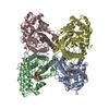
 Components Components | Fructose-bisphosphate aldolase A | ||||||
 Keywords Keywords | LYASE / aldolase / mutant / iminium / Schiff base / intermediate / covalent / Acetylation / Glycolysis / Phosphoprotein | ||||||
| Function / homology |  Function and homology information Function and homology informationnegative regulation of Arp2/3 complex-mediated actin nucleation / fructose-bisphosphate aldolase / fructose-bisphosphate aldolase activity / M band / I band / glycolytic process / protein homotetramerization / positive regulation of cell migration Similarity search - Function | ||||||
| Biological species |  | ||||||
| Method |  X-RAY DIFFRACTION / SYNCHROTRON / MOLECULAR REPLACEMENT / Resolution: 2.03 Å X-RAY DIFFRACTION / SYNCHROTRON / MOLECULAR REPLACEMENT / Resolution: 2.03 Å | ||||||
 Authors Authors | St-Jean, M. / Sygusch, J. | ||||||
 Citation Citation | Journal: Biochemistry / Year: 2009 Title: Charge stabilization and entropy reduction of central lysine residues in fructose-bisphosphate aldolase Authors: St-Jean, M. / Blonski, C. / Sygusch, J. | ||||||
| History |
|
- Structure visualization
| Structure viewer | Molecule: MolmilJmol/JSmol |
|---|
- Downloads & links
-Download
| PDBx/mmCIF format | 3dfs.cif.gz | 326 KB | Display | PDBx/mmCIF format |
|---|---|---|---|---|
| PDB format | pdb3dfs.ent.gz | 259.1 KB | Display | PDB format |
| PDBx/mmJSON format | 3dfs.json.gz | Tree view | PDBx/mmJSON format | |
| Others |  Other downloads Other downloads |
-Validation report
| Arichive directory | https://data.pdbj.org/pub/pdb/validation_reports/df/3dfsftp://data.pdbj.org/pub/pdb/validation_reports/df/3dfs | HTTPS FTP |
|---|
-Related structure data
| Related structure data |  3dfnC 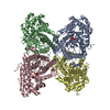 3dfoC 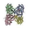 3dfpC  3dfqC  3dftC 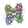 1zahS C: citing same article ( S: Starting model for refinement |
|---|---|
| Similar structure data |


-Links
 PDBj
PDBj
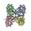
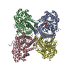
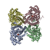
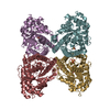
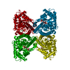
- Assembly
| Deposited unit | 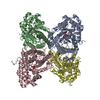
| ||||||||
|---|---|---|---|---|---|---|---|---|---|
| 1 |
| ||||||||
| Unit cell |
|
-Components
| #1: Protein | Mass: 39235.660 Da / Num. of mol.: 4 / Mutation: D33S Source method: isolated from a genetically manipulated source Source: (gene. exp.)  #2: Chemical | ChemComp-13P / 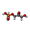  Mass: 170.058 Da / Num. of mol.: 4 / Source method: obtained synthetically / Formula: C3H7O6P Mass: 170.058 Da / Num. of mol.: 4 / Source method: obtained synthetically / Formula: C3H7O6P#3: Water | ChemComp-HOH / |  Mass: 18.015 Da / Num. of mol.: 2120 / Source method: isolated from a natural source / Formula: H2O Mass: 18.015 Da / Num. of mol.: 2120 / Source method: isolated from a natural source / Formula: H2OHas protein modification | Y | Nonpolymer details | 13P LIGAND IS COVALENTLY | |
|---|
-Experimental details
-Experiment
| Experiment | Method: X-RAY DIFFRACTION / Number of used crystals: 1 |
|---|
- Sample preparation
| Crystal | Density Matthews: 2.32 Å3/Da / Density % sol: 47.08 % |
|---|---|
| Crystal grow | Temperature: 296 K / Method: vapor diffusion, hanging drop / pH: 7.5 Details: sodium HEPES, PEG 4000, pH 7.5, VAPOR DIFFUSION, HANGING DROP, temperature 296K |
-Data collection
| Diffraction | Mean temperature: 100 K |
|---|---|
| Diffraction source | Source: SYNCHROTRON / Site: NSLS  / Beamline: X8C / Wavelength: 1.1 Å / Beamline: X8C / Wavelength: 1.1 Å |
| Detector | Type: ADSC QUANTUM 4 / Detector: CCD / Date: Mar 15, 2006 |
| Radiation | Protocol: SINGLE WAVELENGTH / Monochromatic (M) / Laue (L): M / Scattering type: x-ray |
| Radiation wavelength | Wavelength: 1.1 Å / Relative weight: 1 |
| Reflection | Resolution: 1.8→50 Å / Num. obs: 109998 / % possible obs: 82.4 % / Redundancy: 3.6 % / Rsym value: 0.065 / Net I/σ(I): 19.1 |
| Reflection shell | Resolution: 1.8→1.86 Å / Redundancy: 2.8 % / Mean I/σ(I) obs: 2.1 / Num. unique all: 4669 / Rsym value: 0.528 / % possible all: 35.1 |
- Processing
| Software |
| |||||||||||||||||||||||||
|---|---|---|---|---|---|---|---|---|---|---|---|---|---|---|---|---|---|---|---|---|---|---|---|---|---|---|
| Refinement | Method to determine structure: MOLECULAR REPLACEMENT Starting model: PDB entry 1ZAH Resolution: 2.03→44.11 Å / Isotropic thermal model: anisotropic / Cross valid method: THROUGHOUT / σ(I): 1
| |||||||||||||||||||||||||
| Displacement parameters | Biso mean: 20.5 Å2
| |||||||||||||||||||||||||
| Refine analyze |
| |||||||||||||||||||||||||
| Refinement step | Cycle: LAST / Resolution: 2.03→44.11 Å
| |||||||||||||||||||||||||
| Refine LS restraints |
| |||||||||||||||||||||||||
| LS refinement shell | Resolution: 2.03→2.13 Å
|
