[English] 日本語
Yorodumi
- PDB-2quu: Dihydroxyacetone phosphate Schiff base intermediate in mutant fru... -

+
Open data


ID or keywords:

Loading...

-
Basic information

Entry
Database: PDB / ID: 2quu
TitleDihydroxyacetone phosphate Schiff base intermediate in mutant fructose-1,6-bisphosphate aldolase from rabbit muscle
ComponentsFructose-bisphosphate aldolase A
KeywordsLYASE / aldolase / mutant / substrate / Schiff base / protonated imine / intermediate / covalent / Acetylation / Glycolysis / Phosphorylation
Function / homology
Function and homology information


negative regulation of Arp2/3 complex-mediated actin nucleation / fructose-bisphosphate aldolase / fructose-bisphosphate aldolase activity / M band / I band / glycolytic process / protein homotetramerization / positive regulation of cell migration
Similarity search - Function
Fructose-bisphosphate aldolase class-I active site / Fructose-bisphosphate aldolase class-I active site. / Fructose-bisphosphate aldolase, class-I / Fructose-bisphosphate aldolase class-I / Aldolase class I / Aldolase-type TIM barrel / TIM Barrel / Alpha-Beta Barrel / Alpha Beta
Similarity search - Domain/homology
1,3-DIHYDROXYACETONEPHOSPHATE / Fructose-bisphosphate aldolase A
Similarity search - Component
Biological speciesOryctolagus cuniculus (rabbit)
MethodX-RAY DIFFRACTION / SYNCHROTRON / MOLECULAR REPLACEMENT / Resolution: 1.98 Å
AuthorsSt-Jean, M. / Sygusch, J.
CitationJournal: J.Biol.Chem. / Year: 2007
Title: Stereospecific proton transfer by a mobile catalyst in mammalian fructose-1,6-bisphosphate aldolase
Authors: St-Jean, M. / Sygusch, J.
History
DepositionAug 6, 2007Deposition site: RCSB / Processing site: RCSB
Revision 1.0Aug 28, 2007Provider: repository / Type: Initial release
Revision 1.1Jul 13, 2011Group: Version format compliance
Revision 1.2Oct 25, 2017Group: Refinement description / Category: software
Revision 1.3Oct 20, 2021Group: Database references / Derived calculations
Category: database_2 / struct_conn ...database_2 / struct_conn / struct_ref_seq_dif / struct_site
Item: _database_2.pdbx_DOI / _database_2.pdbx_database_accession ..._database_2.pdbx_DOI / _database_2.pdbx_database_accession / _struct_conn.pdbx_leaving_atom_flag / _struct_ref_seq_dif.details / _struct_site.pdbx_auth_asym_id / _struct_site.pdbx_auth_comp_id / _struct_site.pdbx_auth_seq_id
Revision 1.4Aug 30, 2023Group: Data collection / Refinement description
Category: chem_comp_atom / chem_comp_bond / pdbx_initial_refinement_model
Remark 600 HETEROGEN 13P LIGAND IS COVALENTLY BOUND TO LYS-229 AS A PROTONATED SCHIFF BASE (IMINIUM) INTERMEDIATE.

-
Structure visualization

Structure viewerMolecule:
MolmilJmol/JSmol

Downloads & links

-
Assembly

Deposited unit
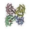
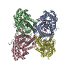
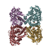
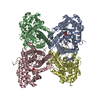
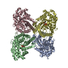
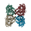
A: Fructose-bisphosphate aldolase A
B: Fructose-bisphosphate aldolase A
C: Fructose-bisphosphate aldolase A
D: Fructose-bisphosphate aldolase A
hetero molecules


Theoretical massNumber of molelcules
Total (without water)157,7438
Polymers157,0634
Non-polymers6804
Water40,1732230
1


  • Idetical with deposited unit
  • defined by author&software
TypeNameSymmetry operationNumber
identity operation1_555x,y,z1
Buried area12420 Å2
MethodPISA
Unit cell
Length a, b, c (Å)83.858, 103.722, 84.881
Angle α, β, γ (deg.)90.000, 98.771, 90.000
Int Tables number4
Space group name H-MP1211
DetailsThe biological assembly is the homotetramer found in the asymmetric unit

-
Components

#1: Protein
Fructose-bisphosphate aldolase A / Muscle-type aldolase


Mass: 39265.688 Da / Num. of mol.: 4 / Mutation: K146M
Source method: isolated from a genetically manipulated source
Source: (gene. exp.) Oryctolagus cuniculus (rabbit) / Gene: ALDOA / Plasmid: pPB14 / Production host: Escherichia coli (E. coli) / Strain (production host): BL-21 SI / References: UniProt: P00883, fructose-bisphosphate aldolase
#2: Chemical
ChemComp-13P / 1,3-DIHYDROXYACETONEPHOSPHATE / Dihydroxyacetone phosphate


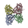
Mass: 170.058 Da / Num. of mol.: 4 / Source method: obtained synthetically / Formula: C3H7O6P
#3: Water ChemComp-HOH / water / Water


Mass: 18.015 Da / Num. of mol.: 2230 / Source method: isolated from a natural source / Formula: H2O
Nonpolymer detailsREMARK 600 HETEROGEN REMARK 600 13P LIGAND IS COVALENTLY BOUND TO LYS-229 AS A SCHIFF BASE ...REMARK 600 HETEROGEN REMARK 600 13P LIGAND IS COVALENTLY BOUND TO LYS-229 AS A SCHIFF BASE INTERMEDIATE (PROTONATED IMINE).

-
Experimental details

-
Experiment

ExperimentMethod: X-RAY DIFFRACTION / Number of used crystals: 1

-
Sample preparation

CrystalDensity Matthews: 2.32 Å3/Da / Density % sol: 47.05 %
Crystal growTemperature: 296 K / Method: vapor diffusion, hanging drop / pH: 7.5
Details: sodium HEPES, PEG 4000, pH 7.5, VAPOR DIFFUSION, HANGING DROP, temperature 296K

-
Data collection

DiffractionMean temperature: 100 K
Diffraction sourceSource: SYNCHROTRON / Site: NSLS / Beamline: X8C / Wavelength: 1.1 Å
DetectorType: ADSC QUANTUM 4 / Detector: CCD / Date: Mar 1, 2006
RadiationProtocol: SINGLE WAVELENGTH / Monochromatic (M) / Laue (L): M / Scattering type: x-ray
Radiation wavelengthWavelength: 1.1 Å / Relative weight: 1
ReflectionResolution: 1.84→50 Å / Num. obs: 109942 / % possible obs: 88.1 % / Redundancy: 3.4 % / Rsym value: 0.052 / Χ2: 1.091 / Net I/σ(I): 21.4
Reflection shellResolution: 1.84→1.91 Å / Num. unique all: 5901 / Χ2: 1.011 / % possible all: 47.5

-
Processing

Software
NameVersionClassificationNB
DENZOdata reduction
SCALEPACKdata scaling
CNS1.1refinement
PDB_EXTRACT2data extraction
CBASSdata collection
HKL-2000data reduction
HKL-2000data scaling
CNSphasing
RefinementMethod to determine structure: MOLECULAR REPLACEMENT
Starting model: PDB ENTRY 1ZAH
Resolution: 1.98→50 Å / Isotropic thermal model: anisotropic / Cross valid method: THROUGHOUT / σ(I): 1
RfactorNum. reflection% reflectionSelection details
Rfree0.195 8893 -random
Rwork0.157 ---
all-96349 --
obs-89297 89.4 %-
Displacement parametersBiso mean: 25.1 Å2
Baniso -1Baniso -2Baniso -3
1-0.402 Å20 Å21.958 Å2
2---5.78 Å20 Å2
3---5.379 Å2
Refine analyze
FreeObs
Luzzati coordinate error0.22 Å0.18 Å
Luzzati d res low-5 Å
Luzzati sigma a0.2 Å0.17 Å
Refinement stepCycle: LAST / Resolution: 1.98→50 Å
ProteinNucleic acidLigandSolventTotal
Num. atoms10820 0 36 2230 13086
Refine LS restraints
Refine-IDTypeDev ideal
X-RAY DIFFRACTIONc_angle_deg1.211
X-RAY DIFFRACTIONc_bond_d0.005
LS refinement shellResolution: 1.98→2.07 Å
RfactorNum. reflection% reflection
Rfree0.248 732 -
Rwork0.213 --
obs-7353 59.2 %

+
About Yorodumi

-
News

-
Feb 9, 2022. New format data for meta-information of EMDB entries

New format data for meta-information of EMDB entries

  • Version 3 of the EMDB header file is now the official format.
  • The previous official version 1.9 will be removed from the archive.

Related info.:EMDB header

External links:wwPDB to switch to version 3 of the EMDB data model

-
Aug 12, 2020. Covid-19 info

Covid-19 info

URL: https://pdbj.org/emnavi/covid19.php

New page: Covid-19 featured information page in EM Navigator.

Related info.:Covid-19 info / Mar 5, 2020. Novel coronavirus structure data

+
Mar 5, 2020. Novel coronavirus structure data

Novel coronavirus structure data

Related info.:Yorodumi Speices / Aug 12, 2020. Covid-19 info

External links:COVID-19 featured content - PDBj / Molecule of the Month (242):Coronavirus Proteases

+
Jan 31, 2019. EMDB accession codes are about to change! (news from PDBe EMDB page)

EMDB accession codes are about to change! (news from PDBe EMDB page)

  • The allocation of 4 digits for EMDB accession codes will soon come to an end. Whilst these codes will remain in use, new EMDB accession codes will include an additional digit and will expand incrementally as the available range of codes is exhausted. The current 4-digit format prefixed with “EMD-” (i.e. EMD-XXXX) will advance to a 5-digit format (i.e. EMD-XXXXX), and so on. It is currently estimated that the 4-digit codes will be depleted around Spring 2019, at which point the 5-digit format will come into force.
  • The EM Navigator/Yorodumi systems omit the EMD- prefix.

Related info.:Q: What is EMD? / ID/Accession-code notation in Yorodumi/EM Navigator

External links:EMDB Accession Codes are Changing Soon! / Contact to PDBj

+
Jul 12, 2017. Major update of PDB

Major update of PDB

  • wwPDB released updated PDB data conforming to the new PDBx/mmCIF dictionary.
  • This is a major update changing the version number from 4 to 5, and with Remediation, in which all the entries are updated.
  • In this update, many items about electron microscopy experimental information are reorganized (e.g. em_software).
  • Now, EM Navigator and Yorodumi are based on the updated data.

External links:wwPDB Remediation / Enriched Model Files Conforming to OneDep Data Standards Now Available in the PDB FTP Archive

-
Yorodumi

Thousand views of thousand structures

  • Yorodumi is a browser for structure data from EMDB, PDB, SASBDB, etc.
  • This page is also the successor to EM Navigator detail page, and also detail information page/front-end page for Omokage search.
  • The word "yorodu" (or yorozu) is an old Japanese word meaning "ten thousand". "mi" (miru) is to see.

Related info.:EMDB / PDB / SASBDB / Comparison of 3 databanks / Yorodumi Search / Aug 31, 2016. New EM Navigator & Yorodumi / Yorodumi Papers / Jmol/JSmol / Function and homology information / Changes in new EM Navigator and Yorodumi

Read more