 Movie
Movie Controller


+ Open data

- Basic information
| Entry | Database: PDB / ID: 1xfb | ||||||
|---|---|---|---|---|---|---|---|
| Title | Human Brain Fructose 1,6-(bis)phosphate Aldolase (C isozyme) | ||||||
 Components Components | Aldolase C | ||||||
 Keywords Keywords | LYASE / isozyme specificity / structural enzymology / protein-protein interactions / isozyme specific residues / structure/function | ||||||
| Function / homology |  Function and homology information Function and homology informationfructose-bisphosphate aldolase / fructose-bisphosphate aldolase activity / fructose 1,6-bisphosphate metabolic process / fructose metabolic process / Gluconeogenesis / Glycolysis / cytoskeletal protein binding / epithelial cell differentiation / glycolytic process / gluconeogenesis ...fructose-bisphosphate aldolase / fructose-bisphosphate aldolase activity / fructose 1,6-bisphosphate metabolic process / fructose metabolic process / Gluconeogenesis / Glycolysis / cytoskeletal protein binding / epithelial cell differentiation / glycolytic process / gluconeogenesis / tertiary granule lumen / secretory granule lumen / ficolin-1-rich granule lumen / cytoskeleton / Neutrophil degranulation / extracellular exosome / extracellular region / cytosol Similarity search - Function | ||||||
| Biological species |  Homo sapiens (human) Homo sapiens (human) | ||||||
| Method |  X-RAY DIFFRACTION / MOLECULAR REPLACEMENT / Resolution: 3 Å X-RAY DIFFRACTION / MOLECULAR REPLACEMENT / Resolution: 3 Å | ||||||
 Authors Authors | Arakaki, T.L. / Pezza, J.A. / Cronin, M.A. / Hopkins, C.E. / Zimmer, D.B. / Tolan, D.R. / Allen, K.N. | ||||||
 Citation Citation | Journal: Protein Sci. / Year: 2004 Title: Structure of human brain fructose 1,6-(bis)phosphate aldolase: linking isozyme structure with function Authors: Arakaki, T.L. / Pezza, J.A. / Cronin, M.A. / Hopkins, C.E. / Zimmer, D.B. / Tolan, D.R. / Allen, K.N. | ||||||
| History |
|
- Structure visualization
| Structure viewer | Molecule: MolmilJmol/JSmol |
|---|
- Downloads & links
-Download
| PDBx/mmCIF format | 1xfb.cif.gz | 678.9 KB | Display | PDBx/mmCIF format |
|---|---|---|---|---|
| PDB format | pdb1xfb.ent.gz | 578.3 KB | Display | PDB format |
| PDBx/mmJSON format | 1xfb.json.gz | Tree view | PDBx/mmJSON format | |
| Others |  Other downloads Other downloads |
-Validation report
| Arichive directory | https://data.pdbj.org/pub/pdb/validation_reports/xf/1xfbftp://data.pdbj.org/pub/pdb/validation_reports/xf/1xfb | HTTPS FTP |
|---|
-Related structure data
| Related structure data | 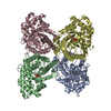 1j4eS S: Starting model for refinement |
|---|---|
| Similar structure data |


-Links
 PDBj
PDBj

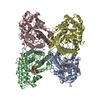
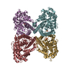
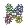
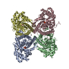
- Assembly
| Deposited unit | 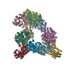
| ||||||||
|---|---|---|---|---|---|---|---|---|---|
| 1 | 
| ||||||||
| 2 | 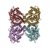
| ||||||||
| 3 | 
| ||||||||
| Unit cell |
| ||||||||
| Details | Tetramer of molecules A, B, C, D / Tetramer of molecules E, F, G, H / Tetramer of molecules I, J, K, L |
-Components
| #1: Protein | Mass: 39616.973 Da / Num. of mol.: 12 Source method: isolated from a genetically manipulated source Source: (gene. exp.) Homo sapiens (human) / Plasmid: pPB1 / Production host:  |
|---|
-Experimental details
-Experiment
| Experiment | Method: X-RAY DIFFRACTION / Number of used crystals: 1 |
|---|
- Sample preparation
| Crystal | Density Matthews: 2.3 Å3/Da / Density % sol: 47 % |
|---|---|
| Crystal grow | Temperature: 291 K / Method: vapor diffusion, hanging drop / pH: 8 Details: PEG 8000, Calcium Acetate, Tris, glucose, pH 8.0, VAPOR DIFFUSION, HANGING DROP, temperature 291K |
-Data collection
| Diffraction | Mean temperature: 93 K |
|---|---|
| Diffraction source | Source: ROTATING ANODE / Type: RIGAKU RU300 / Wavelength: 1.5418 Å |
| Detector | Type: RIGAKU RAXIS IV / Detector: IMAGE PLATE / Date: Feb 1, 2003 / Details: osmic mirrors |
| Radiation | Monochromator: osmic mirrors / Protocol: SINGLE WAVELENGTH / Monochromatic (M) / Laue (L): M / Scattering type: x-ray |
| Radiation wavelength | Wavelength: 1.5418 Å / Relative weight: 1 |
| Reflection | Resolution: 3→79.24 Å / Num. all: 89550 / Num. obs: 89550 / % possible obs: 97.2 % / Observed criterion σ(F): 0 / Observed criterion σ(I): 0 / Limit h max: 27 / Limit h min: -29 / Limit k max: 41 / Limit k min: -29 / Limit l max: 46 / Limit l min: 0 / Observed criterion F max: 1989158.51 / Observed criterion F min: 6.4 / Rsym value: 0.097 / Net I/σ(I): 5.3 |
| Reflection shell | Resolution: 3→3.15 Å / Mean I/σ(I) obs: 1.5 / Rsym value: 0.34 / % possible all: 95.5 |
- Processing
| Software |
| ||||||||||||||||||||||||||||||||||||||||||||||||||||||||||||||||||||||||||||||||||||||||||
|---|---|---|---|---|---|---|---|---|---|---|---|---|---|---|---|---|---|---|---|---|---|---|---|---|---|---|---|---|---|---|---|---|---|---|---|---|---|---|---|---|---|---|---|---|---|---|---|---|---|---|---|---|---|---|---|---|---|---|---|---|---|---|---|---|---|---|---|---|---|---|---|---|---|---|---|---|---|---|---|---|---|---|---|---|---|---|---|---|---|---|---|
| Refinement | Method to determine structure: MOLECULAR REPLACEMENT Starting model: PDB Entry 1J4E Resolution: 3→79.24 Å / Rfactor Rfree error: 0.003 / Occupancy max: 1 / Occupancy min: 1 / Cross valid method: THROUGHOUT / σ(F): 0 / Stereochemistry target values: Engh & Huber
| ||||||||||||||||||||||||||||||||||||||||||||||||||||||||||||||||||||||||||||||||||||||||||
| Solvent computation | Solvent model: CNS bulk solvent model used / Bsol: 30.8274 Å2 / ksol: 0.370575 e/Å3 | ||||||||||||||||||||||||||||||||||||||||||||||||||||||||||||||||||||||||||||||||||||||||||
| Displacement parameters | Biso max: 99.51 Å2 / Biso mean: 35.37 Å2 / Biso min: 0.51 Å2
| ||||||||||||||||||||||||||||||||||||||||||||||||||||||||||||||||||||||||||||||||||||||||||
| Refine analyze |
| ||||||||||||||||||||||||||||||||||||||||||||||||||||||||||||||||||||||||||||||||||||||||||
| Refinement step | Cycle: LAST / Resolution: 3→79.24 Å
| ||||||||||||||||||||||||||||||||||||||||||||||||||||||||||||||||||||||||||||||||||||||||||
| Refine LS restraints |
| ||||||||||||||||||||||||||||||||||||||||||||||||||||||||||||||||||||||||||||||||||||||||||
| LS refinement shell | Refine-ID: X-RAY DIFFRACTION / Total num. of bins used: 8
| ||||||||||||||||||||||||||||||||||||||||||||||||||||||||||||||||||||||||||||||||||||||||||
| Xplor file | Serial no: 1 / Param file: protein_rep.param / Topol file: protein.top |
