 Movie
Movie Controller


+ Open data

- Basic information
| Entry | Database: PDB / ID: 5f3h | ||||||
|---|---|---|---|---|---|---|---|
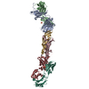
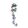
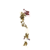
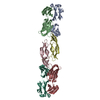
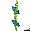
| Title | Structure of myostatin in complex with humanized RK35 antibody | ||||||
 Components Components |
| ||||||
 Keywords Keywords | Signaling Protein/Immune System / myostatin / antibody / complex / Signaling Protein-Immune System complex | ||||||
| Function / homology |  Function and homology information Function and homology informationnegative regulation of muscle hypertrophy / negative regulation of skeletal muscle tissue growth / negative regulation of myoblast proliferation / skeletal muscle satellite cell differentiation / myoblast migration involved in skeletal muscle regeneration / negative regulation of skeletal muscle satellite cell proliferation / skeletal muscle atrophy / ovulation cycle process / FOXO-mediated transcription of cell cycle genes / negative regulation of satellite cell differentiation ...negative regulation of muscle hypertrophy / negative regulation of skeletal muscle tissue growth / negative regulation of myoblast proliferation / skeletal muscle satellite cell differentiation / myoblast migration involved in skeletal muscle regeneration / negative regulation of skeletal muscle satellite cell proliferation / skeletal muscle atrophy / ovulation cycle process / FOXO-mediated transcription of cell cycle genes / negative regulation of satellite cell differentiation / trophoblast cell migration / negative regulation of myoblast differentiation / muscle cell cellular homeostasis / muscle organ development / response to muscle activity / response to testosterone / positive regulation of macrophage chemotaxis / response to gravity / cellular response to dexamethasone stimulus / skeletal muscle tissue development / response to electrical stimulus / positive regulation of lamellipodium assembly / transforming growth factor beta receptor signaling pathway / negative regulation of insulin receptor signaling pathway / cytokine activity / negative regulation of phosphatidylinositol 3-kinase/protein kinase B signal transduction / growth factor activity / response to estrogen / heparin binding / cellular response to hypoxia / response to ethanol / signaling receptor binding / positive regulation of DNA-templated transcription / protein homodimerization activity / : / identical protein binding Similarity search - Function | ||||||
| Biological species |   Homo sapiens (human) Homo sapiens (human) | ||||||
| Method |  X-RAY DIFFRACTION / SYNCHROTRON / MOLECULAR REPLACEMENT / Resolution: 2.7 Å X-RAY DIFFRACTION / SYNCHROTRON / MOLECULAR REPLACEMENT / Resolution: 2.7 Å | ||||||
 Authors Authors | Parris, K.D. / Mosyak, L. | ||||||
 Citation Citation | Journal: Mabs / Year: 2016 Title: Beyond CDR-grafting: Structure-guided humanization of framework and CDR regions of an anti-myostatin antibody. Authors: Apgar, J.R. / Mader, M. / Agostinelli, R. / Benard, S. / Bialek, P. / Johnson, M. / Gao, Y. / Krebs, M. / Owens, J. / Parris, K. / St Andre, M. / Svenson, K. / Morris, C. / Tchistiakova, L. | ||||||
| History |
|
- Structure visualization
| Structure viewer | Molecule: MolmilJmol/JSmol |
|---|
- Downloads & links
-Download
| PDBx/mmCIF format | 5f3h.cif.gz | 394.9 KB | Display | PDBx/mmCIF format |
|---|---|---|---|---|
| PDB format | pdb5f3h.ent.gz | 322.5 KB | Display | PDB format |
| PDBx/mmJSON format | 5f3h.json.gz | Tree view | PDBx/mmJSON format | |
| Others |  Other downloads Other downloads |
-Validation report
| Arichive directory | https://data.pdbj.org/pub/pdb/validation_reports/f3/5f3hftp://data.pdbj.org/pub/pdb/validation_reports/f3/5f3h | HTTPS FTP |
|---|
-Related structure data
| Related structure data |  5f3bSC S: Starting model for refinement C: citing same article ( |
|---|---|
| Similar structure data |


-Links
 PDBj
PDBj


- Assembly
| Deposited unit | 
| ||||||||
|---|---|---|---|---|---|---|---|---|---|
| 1 | 
| ||||||||
| 2 | 
| ||||||||
| Unit cell |
| ||||||||
| Details | This structure contains two biological assemblies. Myostatin is a dimer in solution. Each Myostatin monomer is in complex with RK35 Fab Heavy and Light chains. Thus in this structure one assembly is chains A/B/C/D/I/J the second assembly is E/F/G/H/K/L. |
-Components
| #1: Antibody | Mass: 23369.123 Da / Num. of mol.: 4 Source method: isolated from a genetically manipulated source Source: (gene. exp.)  Cricetulus griseus (Chinese hamster) Cricetulus griseus (Chinese hamster)#2: Antibody | Mass: 23374.879 Da / Num. of mol.: 4 Source method: isolated from a genetically manipulated source Source: (gene. exp.) Cricetulus griseus (Chinese hamster)#3: Protein | Mass: 12307.124 Da / Num. of mol.: 4 Source method: isolated from a genetically manipulated source Source: (gene. exp.) Homo sapiens (human) / Gene: MSTN, GDF8 / Production host: Cricetus (mammal) / References: UniProt: O14793Has protein modification | Y | |
|---|
-Experimental details
-Experiment
| Experiment | Method: X-RAY DIFFRACTION / Number of used crystals: 1 |
|---|
- Sample preparation
| Crystal | Density Matthews: 2.53 Å3/Da / Density % sol: 51.41 % |
|---|---|
| Crystal grow | Temperature: 291 K / Method: vapor diffusion, hanging drop Details: Humanized RK35 Fab and myostatin were purified and the protein complex was concentrated to 10 mg/ml in a buffer of 50 mM tris hydrochloride pH 7.5 and 100 mM sodium chloride. Crystals were ...Details: Humanized RK35 Fab and myostatin were purified and the protein complex was concentrated to 10 mg/ml in a buffer of 50 mM tris hydrochloride pH 7.5 and 100 mM sodium chloride. Crystals were obtained using the hanging drop method with equilibration at 18C against an unbuffered solution containing 20% PEG 3350 and 200mM sodium chloride. |
-Data collection
| Diffraction | Mean temperature: 100 K |
|---|---|
| Diffraction source | Source: SYNCHROTRON / Site: APS  / Beamline: 22-ID / Wavelength: 1 Å / Beamline: 22-ID / Wavelength: 1 Å |
| Detector | Type: ADSC QUANTUM 315 / Detector: CCD / Date: Oct 18, 2010 |
| Radiation | Protocol: SINGLE WAVELENGTH / Monochromatic (M) / Laue (L): M / Scattering type: x-ray |
| Radiation wavelength | Wavelength: 1 Å / Relative weight: 1 |
| Reflection | Resolution: 2.7→50 Å / Num. obs: 40287 |
- Processing
| Software |
| ||||||||||||||||||||
|---|---|---|---|---|---|---|---|---|---|---|---|---|---|---|---|---|---|---|---|---|---|
| Refinement | Method to determine structure: MOLECULAR REPLACEMENT Starting model: 5F3B Resolution: 2.7→19.99 Å / Cor.coef. Fo:Fc: 0.842 / Cor.coef. Fo:Fc free: 0.7929 / Cross valid method: THROUGHOUT / σ(F): 0 / SU Rfree Blow DPI: 0.504
| ||||||||||||||||||||
| Displacement parameters | Biso max: 148.52 Å2 / Biso mean: 39.13 Å2 / Biso min: 3 Å2
| ||||||||||||||||||||
| Refine analyze | Luzzati coordinate error obs: 0.443 Å | ||||||||||||||||||||
| Refinement step | Cycle: final / Resolution: 2.7→19.99 Å
| ||||||||||||||||||||
| LS refinement shell | Resolution: 2.7→2.77 Å / Total num. of bins used: 20
|
