 Movie
Movie Controller


+ Open data

- Basic information
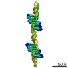
| Entry | Database: PDB / ID: 5f3b | ||||||
|---|---|---|---|---|---|---|---|
| Title | Structure of myostatin in complex with chimeric RK35 antibody | ||||||
 Components Components |
| ||||||
 Keywords Keywords | Signaling Protein/Immune System / myostatin / antibody / complex / Signaling Protein-Immune System complex | ||||||
| Function / homology |  Function and homology information Function and homology informationnegative regulation of muscle hypertrophy / negative regulation of skeletal muscle tissue growth / negative regulation of myoblast proliferation / skeletal muscle satellite cell differentiation / myoblast migration involved in skeletal muscle regeneration / negative regulation of skeletal muscle satellite cell proliferation / skeletal muscle atrophy / ovulation cycle process / FOXO-mediated transcription of cell cycle genes / negative regulation of satellite cell differentiation ...negative regulation of muscle hypertrophy / negative regulation of skeletal muscle tissue growth / negative regulation of myoblast proliferation / skeletal muscle satellite cell differentiation / myoblast migration involved in skeletal muscle regeneration / negative regulation of skeletal muscle satellite cell proliferation / skeletal muscle atrophy / ovulation cycle process / FOXO-mediated transcription of cell cycle genes / negative regulation of satellite cell differentiation / trophoblast cell migration / negative regulation of myoblast differentiation / muscle organ development / muscle cell cellular homeostasis / response to muscle activity / response to testosterone / positive regulation of macrophage chemotaxis / response to gravity / response to electrical stimulus / cellular response to dexamethasone stimulus / skeletal muscle tissue development / positive regulation of lamellipodium assembly / negative regulation of insulin receptor signaling pathway / transforming growth factor beta receptor signaling pathway / cytokine activity / negative regulation of phosphatidylinositol 3-kinase/protein kinase B signal transduction / growth factor activity / response to estrogen / heparin binding / cellular response to hypoxia / response to ethanol / signaling receptor binding / positive regulation of DNA-templated transcription / protein homodimerization activity / : / identical protein binding Similarity search - Function | ||||||
| Biological species |   Homo sapiens (human) Homo sapiens (human) | ||||||
| Method |  X-RAY DIFFRACTION / SYNCHROTRON / MOLECULAR REPLACEMENT / Resolution: 1.76 Å X-RAY DIFFRACTION / SYNCHROTRON / MOLECULAR REPLACEMENT / Resolution: 1.76 Å | ||||||
 Authors Authors | Parris, K.D. / Mosyak, L. | ||||||
 Citation Citation | Journal: Mabs / Year: 2016 Title: Beyond CDR-grafting: Structure-guided humanization of framework and CDR regions of an anti-myostatin antibody. Authors: Apgar, J.R. / Mader, M. / Agostinelli, R. / Benard, S. / Bialek, P. / Johnson, M. / Gao, Y. / Krebs, M. / Owens, J. / Parris, K. / St Andre, M. / Svenson, K. / Morris, C. / Tchistiakova, L. | ||||||
| History |
|
- Structure visualization
| Structure viewer | Molecule: MolmilJmol/JSmol |
|---|
- Downloads & links
-Download
| PDBx/mmCIF format | 5f3b.cif.gz | 245.3 KB | Display | PDBx/mmCIF format |
|---|---|---|---|---|
| PDB format | pdb5f3b.ent.gz | 194.3 KB | Display | PDB format |
| PDBx/mmJSON format | 5f3b.json.gz | Tree view | PDBx/mmJSON format | |
| Others |  Other downloads Other downloads |
-Validation report
| Arichive directory | https://data.pdbj.org/pub/pdb/validation_reports/f3/5f3bftp://data.pdbj.org/pub/pdb/validation_reports/f3/5f3b | HTTPS FTP |
|---|
-Related structure data
| Related structure data |  5f3hC 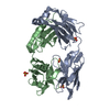 2gcyS S: Starting model for refinement C: citing same article ( |
|---|---|
| Similar structure data |


-Links
 PDBj
PDBj


- Assembly
| Deposited unit | 
| ||||||||
|---|---|---|---|---|---|---|---|---|---|
| 1 |
| ||||||||
| Unit cell |
| ||||||||
| Details | Myostatin exists a dimer and one Fab (heavy/light chain pair) binds to each myostatin, so 6 polymers total. |
-Components
| #1: Antibody | Mass: 23547.418 Da / Num. of mol.: 2 Source method: isolated from a genetically manipulated source Source: (gene. exp.)  Cricetulus griseus (Chinese hamster) Cricetulus griseus (Chinese hamster)#2: Antibody | Mass: 23464.941 Da / Num. of mol.: 2 Source method: isolated from a genetically manipulated source Source: (gene. exp.) Cricetulus griseus (Chinese hamster)#3: Protein | Mass: 12422.212 Da / Num. of mol.: 2 / Fragment: UNP residues 267-375 Source method: isolated from a genetically manipulated source Source: (gene. exp.) Homo sapiens (human) / Gene: MSTN, GDF8 / Production host: Cricetulus griseus (Chinese hamster) / References: UniProt: O14793#4: Chemical | ChemComp-GOL /   Mass: 92.094 Da / Num. of mol.: 5 / Source method: obtained synthetically / Formula: C3H8O3 Mass: 92.094 Da / Num. of mol.: 5 / Source method: obtained synthetically / Formula: C3H8O3#5: Water | ChemComp-HOH / |  Mass: 18.015 Da / Num. of mol.: 1047 / Source method: isolated from a natural source / Formula: H2O Mass: 18.015 Da / Num. of mol.: 1047 / Source method: isolated from a natural source / Formula: H2OHas protein modification | Y | |
|---|
-Experimental details
-Experiment
| Experiment | Method: X-RAY DIFFRACTION / Number of used crystals: 1 |
|---|
- Sample preparation
| Crystal | Density Matthews: 2.67 Å3/Da / Density % sol: 53.97 % |
|---|---|
| Crystal grow | Temperature: 291 K / Method: vapor diffusion, hanging drop / pH: 7.5 Details: Chimeric RK35 Fab and myostatin were purified and the protein complex was concentrated to 10.75 mg/ml in a buffer of 50 mM tris hydrochloride pH 7.5 and 100 mM sodium chloride. Crystals were ...Details: Chimeric RK35 Fab and myostatin were purified and the protein complex was concentrated to 10.75 mg/ml in a buffer of 50 mM tris hydrochloride pH 7.5 and 100 mM sodium chloride. Crystals were obtained using the hanging drop method with equilibration at 18C against a solution containing 20% PEG MME 5000 and 100 mM bis-tris pH 6.5 |
-Data collection
| Diffraction | Mean temperature: 93 K |
|---|---|
| Diffraction source | Source: SYNCHROTRON / Site: APS  / Beamline: 22-ID / Wavelength: 1 Å / Beamline: 22-ID / Wavelength: 1 Å |
| Detector | Type: ADSC QUANTUM 315 / Detector: CCD / Date: Aug 18, 2010 |
| Radiation | Protocol: SINGLE WAVELENGTH / Monochromatic (M) / Laue (L): M / Scattering type: x-ray |
| Radiation wavelength | Wavelength: 1 Å / Relative weight: 1 |
| Reflection | Resolution: 1.76→100 Å / Num. obs: 104791 / Redundancy: 1 % |
- Processing
| Software |
| ||||||||||||||||||||||||
|---|---|---|---|---|---|---|---|---|---|---|---|---|---|---|---|---|---|---|---|---|---|---|---|---|---|
| Refinement | Method to determine structure: MOLECULAR REPLACEMENT Starting model: 2GCY Resolution: 1.76→49.17 Å / Cor.coef. Fo:Fc: 0.9406 / Cor.coef. Fo:Fc free: 0.9276 / SU R Cruickshank DPI: 0.125 / Cross valid method: THROUGHOUT / σ(F): 0 / SU R Blow DPI: 0.129 / SU Rfree Blow DPI: 0.116 / SU Rfree Cruickshank DPI: 0.114
| ||||||||||||||||||||||||
| Displacement parameters | Biso max: 121.64 Å2 / Biso mean: 27.43 Å2 / Biso min: 9.19 Å2
| ||||||||||||||||||||||||
| Refine analyze | Luzzati coordinate error obs: 0.204 Å | ||||||||||||||||||||||||
| Refinement step | Cycle: final / Resolution: 1.76→49.17 Å
| ||||||||||||||||||||||||
| LS refinement shell | Resolution: 1.76→1.81 Å / Total num. of bins used: 20
|
