 Movie
Movie Controller

[English] 日本語
 Yorodumi
- PDB-5ew2: Human thrombin sandwiched between two DNA aptamers: HD22 and HD1-... -
+ Open data

- Basic information
| Entry | Database: PDB / ID: 5ew2 | ||||||
|---|---|---|---|---|---|---|---|
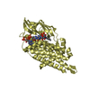
| Title | Human thrombin sandwiched between two DNA aptamers: HD22 and HD1-deltaT12 | ||||||
 Components Components |
| ||||||
 Keywords Keywords | protein/dna / PROTEIN-DNA COMPLEX / BLOOD COAGULATION / DNA APTAMER / DNA-INHIBITOR / G-QUADRUPLEX / DUPLEX-QUADRUPLEX JUNCTION / SERINE PROTEASE / HYDROLASE / ABASIC FURAN / HYDROLASE-DNA COMPLEX | ||||||
| Function / homology |  Function and homology information Function and homology information: / thrombospondin receptor activity / thrombin / thrombin-activated receptor signaling pathway / Defective factor XII causes hereditary angioedema / negative regulation of astrocyte differentiation / regulation of blood coagulation / neutrophil-mediated killing of gram-negative bacterium / positive regulation of phospholipase C-activating G protein-coupled receptor signaling pathway / Defective F8 cleavage by thrombin ...: / thrombospondin receptor activity / thrombin / thrombin-activated receptor signaling pathway / Defective factor XII causes hereditary angioedema / negative regulation of astrocyte differentiation / regulation of blood coagulation / neutrophil-mediated killing of gram-negative bacterium / positive regulation of phospholipase C-activating G protein-coupled receptor signaling pathway / Defective F8 cleavage by thrombin / ligand-gated ion channel signaling pathway / Platelet Aggregation (Plug Formation) / positive regulation of collagen biosynthetic process / negative regulation of platelet activation / negative regulation of blood coagulation / negative regulation of fibrinolysis / blood coagulation, fibrin clot formation / positive regulation of blood coagulation / Transport of gamma-carboxylated protein precursors from the endoplasmic reticulum to the Golgi apparatus / : / Gamma-carboxylation of protein precursors / Removal of aminoterminal propeptides from gamma-carboxylated proteins / regulation of cytosolic calcium ion concentration / fibrinolysis / : / negative regulation of proteolysis / negative regulation of cytokine production involved in inflammatory response / Regulation of Complement cascade / acute-phase response / Cell surface interactions at the vascular wall / positive regulation of release of sequestered calcium ion into cytosol / Peptide ligand-binding receptors / growth factor activity / positive regulation of receptor signaling pathway via JAK-STAT / lipopolysaccharide binding / platelet activation / response to wounding / positive regulation of protein localization to nucleus / Golgi lumen / Regulation of Insulin-like Growth Factor (IGF) transport and uptake by Insulin-like Growth Factor Binding Proteins (IGFBPs) / positive regulation of reactive oxygen species metabolic process / blood coagulation / positive regulation of insulin secretion / regulation of cell shape / antimicrobial humoral immune response mediated by antimicrobial peptide / heparin binding / Thrombin signalling through proteinase activated receptors (PARs) / positive regulation of cell growth / blood microparticle / G alpha (q) signalling events / cell surface receptor signaling pathway / positive regulation of phosphatidylinositol 3-kinase/protein kinase B signal transduction / endoplasmic reticulum lumen / receptor ligand activity / signaling receptor binding / serine-type endopeptidase activity / calcium ion binding / positive regulation of cell population proliferation / proteolysis / : / extracellular exosome / extracellular region / plasma membrane Similarity search - Function | ||||||
| Biological species |  Homo sapiens (human) Homo sapiens (human) | ||||||
| Method |  X-RAY DIFFRACTION / MOLECULAR REPLACEMENT / molecular replacement / Resolution: 3.59 Å X-RAY DIFFRACTION / MOLECULAR REPLACEMENT / molecular replacement / Resolution: 3.59 Å | ||||||
 Authors Authors | Pica, A. / Russo Krauss, I. / Parente, V. / Sica, F. | ||||||
 Citation Citation | Journal: Nucleic Acids Res. / Year: 2017 Title: Through-bond effects in the ternary complexes of thrombin sandwiched by two DNA aptamers. Authors: Pica, A. / Russo Krauss, I. / Parente, V. / Tateishi-Karimata, H. / Nagatoishi, S. / Tsumoto, K. / Sugimoto, N. / Sica, F. #1: Journal: Acta Crystallogr. D Biol. Crystallogr. / Year: 2013Title: Duplex-quadruplex motifs in a peculiar structural organization cooperatively contribute to thrombin binding of a DNA aptamer. Authors: Russo Krauss, I. / Pica, A. / Merlino, A. / Mazzarella, L. / Sica, F. #2: Journal: FEBS J. / Year: 2013Title: Dissecting the contribution of thrombin exosite I in the recognition of thrombin binding aptamer. Authors: Pica, A. / Russo Krauss, I. / Merlino, A. / Nagatoishi, S. / Sugimoto, N. / Sica, F. | ||||||
| History |
|
- Structure visualization
| Structure viewer | Molecule: MolmilJmol/JSmol |
|---|
- Downloads & links
-Download
| PDBx/mmCIF format | 5ew2.cif.gz | 176.6 KB | Display | PDBx/mmCIF format |
|---|---|---|---|---|
| PDB format | pdb5ew2.ent.gz | 135.9 KB | Display | PDB format |
| PDBx/mmJSON format | 5ew2.json.gz | Tree view | PDBx/mmJSON format | |
| Others |  Other downloads Other downloads |
-Validation report
| Arichive directory | https://data.pdbj.org/pub/pdb/validation_reports/ew/5ew2ftp://data.pdbj.org/pub/pdb/validation_reports/ew/5ew2 | HTTPS FTP |
|---|
-Related structure data


-Links
 PDBj
PDBj


- Assembly
| Deposited unit | 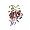
| ||||||||
|---|---|---|---|---|---|---|---|---|---|
| 1 |
| ||||||||
| Unit cell |
|
-Components
-DNA chain , 2 types, 2 molecules DE
| #3: DNA chain | Mass: 8485.429 Da / Num. of mol.: 1 / Source method: obtained synthetically / Source: (synth.) Homo sapiens (human) |
|---|---|
| #4: DNA chain | Mass: 4618.955 Da / Num. of mol.: 1 / Source method: obtained synthetically Details: HD1 variant in which nucleobase of T12 has been deleted Source: (synth.) Homo sapiens (human) |
-Protein/peptide / Protein / Sugars , 3 types, 3 molecules LH
| #1: Protein/peptide | Mass: 4096.534 Da / Num. of mol.: 1 / Source method: isolated from a natural source / Source: (natural) Homo sapiens (human) / References: UniProt: P00734, thrombin |
|---|---|
| #2: Protein | Mass: 29780.219 Da / Num. of mol.: 1 / Source method: isolated from a natural source / Source: (natural) Homo sapiens (human) / References: UniProt: P00734, thrombin |
| #5: Sugar | ChemComp-NAG /  Type: D-saccharide, beta linking / Mass: 221.208 Da / Num. of mol.: 1 Type: D-saccharide, beta linking / Mass: 221.208 Da / Num. of mol.: 1Source method: isolated from a genetically manipulated source Formula: C8H15NO6 |
-Non-polymers , 3 types, 11 molecules 


| #6: Chemical | ChemComp-0G6 /  Type: peptide-like, Peptide-like / Class: Inhibitor / Mass: 453.986 Da / Num. of mol.: 1 / Source method: obtained synthetically / Formula: C21H34ClN6O3 / References: D-Phe-Pro-Arg-CH2Cl Type: peptide-like, Peptide-like / Class: Inhibitor / Mass: 453.986 Da / Num. of mol.: 1 / Source method: obtained synthetically / Formula: C21H34ClN6O3 / References: D-Phe-Pro-Arg-CH2Cl | ||
|---|---|---|---|
| #7: Chemical |  Mass: 22.990 Da / Num. of mol.: 3 / Source method: obtained synthetically / Formula: Na Mass: 22.990 Da / Num. of mol.: 3 / Source method: obtained synthetically / Formula: Na#8: Water | ChemComp-HOH / | Mass: 18.015 Da / Num. of mol.: 7 / Source method: isolated from a natural source / Formula: H2O |
-Details
| Has protein modification | Y |
|---|
-Experimental details
-Experiment
| Experiment | Method: X-RAY DIFFRACTION / Number of used crystals: 1 |
|---|
- Sample preparation
| Crystal | Density Matthews: 2.63 Å3/Da / Density % sol: 53.24 % |
|---|---|
| Crystal grow | Temperature: 298 K / Method: vapor diffusion, hanging drop Details: 18% PEG 3350, 0.2 M Sodium Formate, 2% acetonitrile |
-Data collection
| Diffraction | Mean temperature: 100 K | |||||||||||||||||||||||||||||||||||||||||||||||||||||||||||||||||||||||||||||||||||||||||||||||||||
|---|---|---|---|---|---|---|---|---|---|---|---|---|---|---|---|---|---|---|---|---|---|---|---|---|---|---|---|---|---|---|---|---|---|---|---|---|---|---|---|---|---|---|---|---|---|---|---|---|---|---|---|---|---|---|---|---|---|---|---|---|---|---|---|---|---|---|---|---|---|---|---|---|---|---|---|---|---|---|---|---|---|---|---|---|---|---|---|---|---|---|---|---|---|---|---|---|---|---|---|---|
| Diffraction source | Source: ROTATING ANODE / Type: RIGAKU MICROMAX-007 HF / Wavelength: 1.5418 Å | |||||||||||||||||||||||||||||||||||||||||||||||||||||||||||||||||||||||||||||||||||||||||||||||||||
| Detector | Type: RIGAKU SATURN 944 / Detector: CCD / Date: Nov 13, 2013 | |||||||||||||||||||||||||||||||||||||||||||||||||||||||||||||||||||||||||||||||||||||||||||||||||||
| Radiation | Protocol: SINGLE WAVELENGTH / Monochromatic (M) / Laue (L): M / Scattering type: x-ray | |||||||||||||||||||||||||||||||||||||||||||||||||||||||||||||||||||||||||||||||||||||||||||||||||||
| Radiation wavelength | Wavelength: 1.5418 Å / Relative weight: 1 | |||||||||||||||||||||||||||||||||||||||||||||||||||||||||||||||||||||||||||||||||||||||||||||||||||
| Reflection | Resolution: 3.58→50 Å / Num. obs: 5755 / % possible obs: 96.4 % / Redundancy: 2.8 % / Rmerge(I) obs: 0.12 / Rpim(I) all: 0.084 / Rrim(I) all: 0.147 / Χ2: 0.811 / Net I/av σ(I): 7.51 / Net I/σ(I): 5.9 / Num. measured all: 16010 | |||||||||||||||||||||||||||||||||||||||||||||||||||||||||||||||||||||||||||||||||||||||||||||||||||
| Reflection shell | Diffraction-ID: 1 / Rejects: _
|
-Phasing
| Phasing | Method: molecular replacement | |||||||||
|---|---|---|---|---|---|---|---|---|---|---|
| Phasing MR | Model details: Phaser MODE: MR_AUTO
|
- Processing
| Software |
| ||||||||||||||||||||||||||||||||||||||||||||||||||||||||||||||||||||||||||||||||||||||||||||||||||||||||||||||||||||||||||||||||||||||||||||||||||||||||||||||||||||||||||||||||||||||
|---|---|---|---|---|---|---|---|---|---|---|---|---|---|---|---|---|---|---|---|---|---|---|---|---|---|---|---|---|---|---|---|---|---|---|---|---|---|---|---|---|---|---|---|---|---|---|---|---|---|---|---|---|---|---|---|---|---|---|---|---|---|---|---|---|---|---|---|---|---|---|---|---|---|---|---|---|---|---|---|---|---|---|---|---|---|---|---|---|---|---|---|---|---|---|---|---|---|---|---|---|---|---|---|---|---|---|---|---|---|---|---|---|---|---|---|---|---|---|---|---|---|---|---|---|---|---|---|---|---|---|---|---|---|---|---|---|---|---|---|---|---|---|---|---|---|---|---|---|---|---|---|---|---|---|---|---|---|---|---|---|---|---|---|---|---|---|---|---|---|---|---|---|---|---|---|---|---|---|---|---|---|---|---|
| Refinement | Method to determine structure: MOLECULAR REPLACEMENT / Resolution: 3.59→50 Å / Cor.coef. Fo:Fc: 0.895 / Cor.coef. Fo:Fc free: 0.859 / SU ML: 0.57 / Cross valid method: THROUGHOUT / Stereochemistry target values: MAXIMUM LIKELIHOOD / Details: HYDROGENS HAVE BEEN ADDED IN THE RIDING POSITIONS
| ||||||||||||||||||||||||||||||||||||||||||||||||||||||||||||||||||||||||||||||||||||||||||||||||||||||||||||||||||||||||||||||||||||||||||||||||||||||||||||||||||||||||||||||||||||||
| Solvent computation | Ion probe radii: 0.8 Å / Shrinkage radii: 0.8 Å / VDW probe radii: 1.2 Å / Solvent model: MASK | ||||||||||||||||||||||||||||||||||||||||||||||||||||||||||||||||||||||||||||||||||||||||||||||||||||||||||||||||||||||||||||||||||||||||||||||||||||||||||||||||||||||||||||||||||||||
| Displacement parameters | Biso mean: 70 Å2
| ||||||||||||||||||||||||||||||||||||||||||||||||||||||||||||||||||||||||||||||||||||||||||||||||||||||||||||||||||||||||||||||||||||||||||||||||||||||||||||||||||||||||||||||||||||||
| Refinement step | Cycle: LAST / Resolution: 3.59→50 Å
| ||||||||||||||||||||||||||||||||||||||||||||||||||||||||||||||||||||||||||||||||||||||||||||||||||||||||||||||||||||||||||||||||||||||||||||||||||||||||||||||||||||||||||||||||||||||
| Refine LS restraints |
|