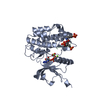
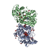
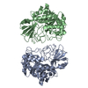
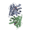
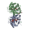
[English] 日本語
Yorodumi
- PDB-4xlv: Crystal structure of the activated insulin receptor tyrosine kina... -

+
Open data


ID or keywords:

Loading...

-
Basic information

Entry
Database: PDB / ID: 4xlv
TitleCrystal structure of the activated insulin receptor tyrosine kinase dimer
ComponentsInsulin receptor
KeywordsTRANSFERASE / TYROSINE KINASE / SIGNAL TRANSDUCTION / PHOSPHORYL TRANSFER
Function / homology
Function and homology information


regulation of female gonad development / positive regulation of meiotic cell cycle / positive regulation of developmental growth / insulin-like growth factor II binding / male sex determination / exocrine pancreas development / insulin receptor complex / insulin-like growth factor I binding / insulin receptor activity / positive regulation of protein-containing complex disassembly ...regulation of female gonad development / positive regulation of meiotic cell cycle / positive regulation of developmental growth / insulin-like growth factor II binding / male sex determination / exocrine pancreas development / insulin receptor complex / insulin-like growth factor I binding / insulin receptor activity / positive regulation of protein-containing complex disassembly / cargo receptor activity / dendritic spine maintenance / insulin binding / PTB domain binding / adrenal gland development / neuronal cell body membrane / Signaling by Insulin receptor / IRS activation / activation of protein kinase activity / amyloid-beta clearance / positive regulation of respiratory burst / positive regulation of receptor internalization / regulation of embryonic development / transport across blood-brain barrier / insulin receptor substrate binding / positive regulation of glycogen biosynthetic process / epidermis development / Signal attenuation / phosphatidylinositol 3-kinase binding / heart morphogenesis / dendrite membrane / Insulin receptor recycling / neuron projection maintenance / positive regulation of glycolytic process / activation of protein kinase B activity / positive regulation of mitotic nuclear division / Insulin receptor signalling cascade / receptor-mediated endocytosis / learning / caveola / positive regulation of glucose import / insulin-like growth factor receptor binding / positive regulation of MAP kinase activity / receptor internalization / receptor protein-tyrosine kinase / memory / cellular response to growth factor stimulus / peptidyl-tyrosine phosphorylation / cellular response to insulin stimulus / male gonad development / positive regulation of nitric oxide biosynthetic process / late endosome / insulin receptor signaling pathway / glucose homeostasis / amyloid-beta binding / PI5P, PP2A and IER3 Regulate PI3K/AKT Signaling / protein tyrosine kinase activity / positive regulation of MAPK cascade / protein autophosphorylation / positive regulation of phosphatidylinositol 3-kinase/protein kinase B signal transduction / lysosome / receptor complex / endosome membrane / positive regulation of cell migration / symbiont entry into host cell / positive regulation of protein phosphorylation / G protein-coupled receptor signaling pathway / protein domain specific binding / external side of plasma membrane / axon / protein phosphorylation / positive regulation of cell population proliferation / protein-containing complex binding / regulation of DNA-templated transcription / GTP binding / positive regulation of DNA-templated transcription / extracellular exosome / ATP binding / identical protein binding / membrane / plasma membrane
Similarity search - Function
Insulin receptor, trans-membrane domain / Insulin receptor trans-membrane segment / Tyrosine-protein kinase, insulin-like receptor / Tyrosine-protein kinase, receptor class II, conserved site / Receptor tyrosine kinase class II signature. / Receptor L-domain / Furin-like cysteine-rich domain / Receptor L-domain superfamily / Furin-like cysteine rich region / Receptor L domain ...Insulin receptor, trans-membrane domain / Insulin receptor trans-membrane segment / Tyrosine-protein kinase, insulin-like receptor / Tyrosine-protein kinase, receptor class II, conserved site / Receptor tyrosine kinase class II signature. / Receptor L-domain / Furin-like cysteine-rich domain / Receptor L-domain superfamily / Furin-like cysteine rich region / Receptor L domain / Furin-like repeat / Furin-like repeats / Growth factor receptor cysteine-rich domain superfamily / Fibronectin type III domain / Fibronectin type 3 domain / Fibronectin type-III domain profile. / Fibronectin type III / Fibronectin type III superfamily / Tyrosine-protein kinase, catalytic domain / Tyrosine kinase, catalytic domain / Tyrosine protein kinases specific active-site signature. / Tyrosine-protein kinase, active site / Protein tyrosine and serine/threonine kinase / Serine-threonine/tyrosine-protein kinase, catalytic domain / Transferase(Phosphotransferase) domain 1 / Transferase(Phosphotransferase); domain 1 / Phosphorylase Kinase; domain 1 / Phosphorylase Kinase; domain 1 / Protein kinase, ATP binding site / Protein kinases ATP-binding region signature. / Immunoglobulin-like fold / Protein kinase domain profile. / Protein kinase domain / Protein kinase-like domain superfamily / 2-Layer Sandwich / Orthogonal Bundle / Mainly Alpha / Alpha Beta
Similarity search - Domain/homology
PHOSPHOMETHYLPHOSPHONIC ACID ADENYLATE ESTER / Insulin receptor
Similarity search - Component
Biological speciesHomo sapiens (human)
MethodX-RAY DIFFRACTION / SYNCHROTRON / Resolution: 2.3 Å
AuthorsHubbard, S.R. / Li, S.
Funding support United States, 1items
OrganizationGrant numberCountry
National Institutes of Health/National Institute of Diabetes and Digestive and Kidney Disease (NIH/NIDDK)DK052916 United States
CitationJournal: Nat Commun / Year: 2015
Title: The insulin and IGF1 receptor kinase domains are functional dimers in the activated state.
Authors: Cabail, M.Z. / Li, S. / Lemmon, E. / Bowen, M.E. / Hubbard, S.R. / Miller, W.T.
History
DepositionJan 13, 2015Deposition site: RCSB / Processing site: RCSB
Revision 1.0Mar 25, 2015Provider: repository / Type: Initial release
Revision 1.1Sep 20, 2017Group: Author supporting evidence / Derived calculations / Source and taxonomy
Category: entity_src_gen / pdbx_audit_support / pdbx_struct_oper_list
Item: _entity_src_gen.pdbx_alt_source_flag / _pdbx_audit_support.funding_organization / _pdbx_struct_oper_list.symmetry_operation
Revision 1.2Dec 25, 2019Group: Author supporting evidence / Category: pdbx_audit_support / Item: _pdbx_audit_support.funding_organization

-
Structure visualization

Structure viewerMolecule:
MolmilJmol/JSmol

Downloads & links

-
Assembly

Deposited unit
A: Insulin receptor
hetero molecules


Theoretical massNumber of molelcules
Total (without water)37,9124
Polymers37,3581
Non-polymers5543
Water2,540141
1
A: Insulin receptor
hetero molecules

A: Insulin receptor
hetero molecules


Theoretical massNumber of molelcules
Total (without water)75,8248
Polymers74,7162
Non-polymers1,1086
Water362
TypeNameSymmetry operationNumber
identity operation1_555x,y,z1
crystal symmetry operation4_556y,x,-z+11
Buried area2610 Å2
ΔGint-15 kcal/mol
Surface area29970 Å2
MethodPISA
Unit cell
Length a, b, c (Å)66.982, 66.982, 136.580
Angle α, β, γ (deg.)90.00, 90.00, 120.00
Int Tables number154
Space group name H-MP3221

-
Components

#1: Protein Insulin receptor / IR


Mass: 37358.008 Da / Num. of mol.: 1 / Fragment: UNP residues 983-1310 / Mutation: Y965F, C981S
Source method: isolated from a genetically manipulated source
Source: (gene. exp.) Homo sapiens (human) / Gene: INSR / Plasmid: pFASTBAC / Production host: 
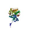
Spodoptera frugiperda (fall armyworm)
References: UniProt: P06213, receptor protein-tyrosine kinase
#2: Chemical ChemComp-ACP / PHOSPHOMETHYLPHOSPHONIC ACID ADENYLATE ESTER / ADENOSINE-5'-[BETA, GAMMA-METHYLENE]TRIPHOSPHATE


Mass: 505.208 Da / Num. of mol.: 1 / Source method: obtained synthetically / Formula: C11H18N5O12P3 / Comment: AMP-PCP, energy-carrying molecule analogue*YM
#3: Chemical ChemComp-MG / MAGNESIUM ION


Mass: 24.305 Da / Num. of mol.: 2 / Source method: obtained synthetically / Formula: Mg
#4: Water ChemComp-HOH / water


Mass: 18.015 Da / Num. of mol.: 141 / Source method: isolated from a natural source / Formula: H2O

-
Experimental details

-
Experiment

ExperimentMethod: X-RAY DIFFRACTION

-
Sample preparation

CrystalDensity Matthews: 2.37 Å3/Da / Density % sol: 48.05 %
Crystal growTemperature: 277 K / Method: vapor diffusion, hanging drop / pH: 7.5
Details: 100 mM Tris-HCl, pH 7.5, 26% (w/v) polyethylene glycol (PEG) 4000, 200 mM MgCl2, and 5 mM DTT

-
Data collection

DiffractionMean temperature: 100 K
Diffraction sourceSource: SYNCHROTRON / Site: NSLS / Beamline: X12C / Wavelength: 0.987 Å
DetectorType: RIGAKU RAXIS IV / Detector: IMAGE PLATE / Date: Apr 15, 2001
RadiationProtocol: SINGLE WAVELENGTH / Monochromatic (M) / Laue (L): M / Scattering type: x-ray
Radiation wavelengthWavelength: 0.987 Å / Relative weight: 1
ReflectionResolution: 2.3→30 Å / Num. obs: 16312 / % possible obs: 98.9 % / Redundancy: 4.9 % / Rsym value: 0.07 / Net I/σ(I): 17.1

-
Processing

Software
NameVersionClassification
REFMAC5.6.0117refinement
HKL-2000data reduction
HKL-2000data scaling
AMoREphasing
RefinementResolution: 2.3→30 Å / Cor.coef. Fo:Fc: 0.934 / Cor.coef. Fo:Fc free: 0.885 / SU B: 7.501 / SU ML: 0.18 / Cross valid method: THROUGHOUT / ESU R: 0.385 / ESU R Free: 0.268 / Stereochemistry target values: MAXIMUM LIKELIHOOD / Details: HYDROGENS HAVE BEEN USED IF PRESENT IN THE INPUT
RfactorNum. reflection% reflectionSelection details
Rfree0.26625 789 5.1 %RANDOM
Rwork0.20422 ---
obs0.20728 14682 94.37 %-
Solvent computationIon probe radii: 0.8 Å / Shrinkage radii: 0.8 Å / VDW probe radii: 1.2 Å / Solvent model: MASK
Displacement parametersBiso mean: 32.319 Å2
Baniso -1Baniso -2Baniso -3
1--1.03 Å2-0.51 Å20 Å2
2---1.03 Å20 Å2
3---1.54 Å2
Refinement stepCycle: LAST / Resolution: 2.3→30 Å
ProteinNucleic acidLigandSolventTotal
Num. atoms2445 0 33 143 2621
Refine LS restraints
Refine-IDTypeDev idealDev ideal targetNumber
X-RAY DIFFRACTIONr_bond_refined_d0.0090.0192535
X-RAY DIFFRACTIONr_bond_other_d
X-RAY DIFFRACTIONr_angle_refined_deg1.4821.9943449
X-RAY DIFFRACTIONr_angle_other_deg
X-RAY DIFFRACTIONr_dihedral_angle_1_deg5.9755309
X-RAY DIFFRACTIONr_dihedral_angle_2_deg34.82824.071113
X-RAY DIFFRACTIONr_dihedral_angle_3_deg15.55315414
X-RAY DIFFRACTIONr_dihedral_angle_4_deg17.2681517
X-RAY DIFFRACTIONr_chiral_restr0.0890.2372
X-RAY DIFFRACTIONr_gen_planes_refined0.0070.0211921
X-RAY DIFFRACTIONr_gen_planes_other
X-RAY DIFFRACTIONr_nbd_refined
X-RAY DIFFRACTIONr_nbd_other
X-RAY DIFFRACTIONr_nbtor_refined
X-RAY DIFFRACTIONr_nbtor_other
X-RAY DIFFRACTIONr_xyhbond_nbd_refined
X-RAY DIFFRACTIONr_xyhbond_nbd_other
X-RAY DIFFRACTIONr_metal_ion_refined
X-RAY DIFFRACTIONr_metal_ion_other
X-RAY DIFFRACTIONr_symmetry_vdw_refined
X-RAY DIFFRACTIONr_symmetry_vdw_other
X-RAY DIFFRACTIONr_symmetry_hbond_refined
X-RAY DIFFRACTIONr_symmetry_hbond_other
X-RAY DIFFRACTIONr_symmetry_metal_ion_refined
X-RAY DIFFRACTIONr_symmetry_metal_ion_other
X-RAY DIFFRACTIONr_mcbond_it
X-RAY DIFFRACTIONr_mcbond_other
X-RAY DIFFRACTIONr_mcangle_it
X-RAY DIFFRACTIONr_mcangle_other
X-RAY DIFFRACTIONr_scbond_it
X-RAY DIFFRACTIONr_scbond_other
X-RAY DIFFRACTIONr_scangle_it
X-RAY DIFFRACTIONr_scangle_other
X-RAY DIFFRACTIONr_long_range_B_refined
X-RAY DIFFRACTIONr_long_range_B_other
X-RAY DIFFRACTIONr_rigid_bond_restr
X-RAY DIFFRACTIONr_sphericity_free
X-RAY DIFFRACTIONr_sphericity_bonded
LS refinement shellResolution: 2.3→2.359 Å / Total num. of bins used: 20
RfactorNum. reflection% reflection
Rfree0.322 43 -
Rwork0.227 884 -
obs--85.12 %

+
About Yorodumi

-
News

-
Feb 9, 2022. New format data for meta-information of EMDB entries

New format data for meta-information of EMDB entries

  • Version 3 of the EMDB header file is now the official format.
  • The previous official version 1.9 will be removed from the archive.

Related info.:EMDB header

External links:wwPDB to switch to version 3 of the EMDB data model

-
Aug 12, 2020. Covid-19 info

Covid-19 info

URL: https://pdbjlvh1.pdbj.org/emnavi/covid19.php

New page: Covid-19 featured information page in EM Navigator.

Related info.:Covid-19 info / Mar 5, 2020. Novel coronavirus structure data

+
Mar 5, 2020. Novel coronavirus structure data

Novel coronavirus structure data

Related info.:Yorodumi Speices / Aug 12, 2020. Covid-19 info

External links:COVID-19 featured content - PDBj / Molecule of the Month (242):Coronavirus Proteases

+
Jan 31, 2019. EMDB accession codes are about to change! (news from PDBe EMDB page)

EMDB accession codes are about to change! (news from PDBe EMDB page)

  • The allocation of 4 digits for EMDB accession codes will soon come to an end. Whilst these codes will remain in use, new EMDB accession codes will include an additional digit and will expand incrementally as the available range of codes is exhausted. The current 4-digit format prefixed with “EMD-” (i.e. EMD-XXXX) will advance to a 5-digit format (i.e. EMD-XXXXX), and so on. It is currently estimated that the 4-digit codes will be depleted around Spring 2019, at which point the 5-digit format will come into force.
  • The EM Navigator/Yorodumi systems omit the EMD- prefix.

Related info.:Q: What is EMD? / ID/Accession-code notation in Yorodumi/EM Navigator

External links:EMDB Accession Codes are Changing Soon! / Contact to PDBj

+
Jul 12, 2017. Major update of PDB

Major update of PDB

  • wwPDB released updated PDB data conforming to the new PDBx/mmCIF dictionary.
  • This is a major update changing the version number from 4 to 5, and with Remediation, in which all the entries are updated.
  • In this update, many items about electron microscopy experimental information are reorganized (e.g. em_software).
  • Now, EM Navigator and Yorodumi are based on the updated data.

External links:wwPDB Remediation / Enriched Model Files Conforming to OneDep Data Standards Now Available in the PDB FTP Archive

-
Yorodumi

Thousand views of thousand structures

  • Yorodumi is a browser for structure data from EMDB, PDB, SASBDB, etc.
  • This page is also the successor to EM Navigator detail page, and also detail information page/front-end page for Omokage search.
  • The word "yorodu" (or yorozu) is an old Japanese word meaning "ten thousand". "mi" (miru) is to see.

Related info.:EMDB / PDB / SASBDB / Comparison of 3 databanks / Yorodumi Search / Aug 31, 2016. New EM Navigator & Yorodumi / Yorodumi Papers / Jmol/JSmol / Function and homology information / Changes in new EM Navigator and Yorodumi

Read more