 Movie
Movie Controller


+ Open data

- Basic information
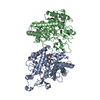
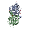
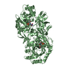
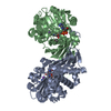
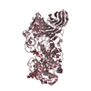
| Entry | Database: PDB / ID: 5xav | ||||||
|---|---|---|---|---|---|---|---|
| Title | Structure of PhaC from Chromobacterium sp. USM2 | ||||||
 Components Components | Intracellular polyhydroxyalkanoate synthase | ||||||
 Keywords Keywords | BIOSYNTHETIC PROTEIN / bioplastic synthase / PhaC | ||||||
| Function / homology | Poly-beta-hydroxybutyrate polymerase, N-terminal domain / Poly(R)-hydroxyalkanoic acid synthase, class I / : / Poly-beta-hydroxybutyrate polymerase (PhaC) N-terminus / poly-hydroxybutyrate biosynthetic process / acyltransferase activity / Alpha/Beta hydrolase fold / cytoplasm / Intracellular polyhydroxyalkanoate synthase Function and homology information Function and homology information | ||||||
| Biological species |  Chromobacterium sp. USM2 (bacteria) Chromobacterium sp. USM2 (bacteria) | ||||||
| Method |  X-RAY DIFFRACTION / SYNCHROTRON / SAD / Resolution: 1.479 Å X-RAY DIFFRACTION / SYNCHROTRON / SAD / Resolution: 1.479 Å | ||||||
 Authors Authors | Chek, M.F. / Kim, S.Y. / Mori, T. / Arsad, H. / Samian, M.R. / Sudesh, K. / Hakoshima, T. | ||||||
| Funding support |  Japan, 1items Japan, 1items
| ||||||
 Citation Citation | Journal: Sci Rep / Year: 2017 Title: Structure of polyhydroxyalkanoate (PHA) synthase PhaC from Chromobacterium sp. USM2, producing biodegradable plastics Authors: Chek, M.F. / Kim, S.Y. / Mori, T. / Arsad, H. / Samian, M.R. / Sudesh, K. / Hakoshima, T. | ||||||
| History |
|
- Structure visualization
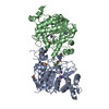
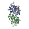
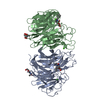
| Structure viewer | Molecule: MolmilJmol/JSmol |
|---|
- Downloads & links
-Download
| PDBx/mmCIF format | 5xav.cif.gz | 348.6 KB | Display | PDBx/mmCIF format |
|---|---|---|---|---|
| PDB format | pdb5xav.ent.gz | 284.9 KB | Display | PDB format |
| PDBx/mmJSON format | 5xav.json.gz | Tree view | PDBx/mmJSON format | |
| Others |  Other downloads Other downloads |
-Validation report
| Arichive directory | https://data.pdbj.org/pub/pdb/validation_reports/xa/5xavftp://data.pdbj.org/pub/pdb/validation_reports/xa/5xav | HTTPS FTP |
|---|
-Related structure data
| Similar structure data |
|---|


-Links
 PDBj
PDBj
- Assembly
| Deposited unit | 
| ||||||||
|---|---|---|---|---|---|---|---|---|---|
| 1 |
| ||||||||
| Unit cell |
|
-Components
| #1: Protein | Mass: 43829.395 Da / Num. of mol.: 2 / Fragment: UNP residues 175-567 Source method: isolated from a genetically manipulated source Source: (gene. exp.) Chromobacterium sp. USM2 (bacteria) / Gene: phaC / Production host: #2: Water | ChemComp-HOH / |  Mass: 18.015 Da / Num. of mol.: 1049 / Source method: isolated from a natural source / Formula: H2O Mass: 18.015 Da / Num. of mol.: 1049 / Source method: isolated from a natural source / Formula: H2O |
|---|
-Experimental details
-Experiment
| Experiment | Method: X-RAY DIFFRACTION / Number of used crystals: 1 |
|---|
- Sample preparation
| Crystal | Density Matthews: 2.4 Å3/Da / Density % sol: 48.77 % |
|---|---|
| Crystal grow | Temperature: 277.15 K / Method: vapor diffusion, hanging drop / pH: 5.5 Details: 50 mM Bis-Tris pH5.5, 60 mM Ammonium sulfate 5% PEG4000 |
-Data collection
| Diffraction | Mean temperature: 100 K |
|---|---|
| Diffraction source | Source: SYNCHROTRON / Site: SPring-8 / Beamline: BL44XU / Wavelength: 0.9 Å |
| Detector | Type: RAYONIX MX300HE / Detector: CCD / Date: Jul 19, 2015 |
| Radiation | Protocol: SINGLE WAVELENGTH / Monochromatic (M) / Laue (L): M / Scattering type: x-ray |
| Radiation wavelength | Wavelength: 0.9 Å / Relative weight: 1 |
| Reflection | Resolution: 1.479→50 Å / Num. obs: 137720 / % possible obs: 99.8 % / Redundancy: 11.2 % / Net I/σ(I): 58.6 |
| Reflection shell | Resolution: 1.48→1.51 Å / % possible obs: 100 % / Redundancy: 11.2 % / Rmerge(I) obs: 0.644 / Mean I/σ(I) obs: 6.2 |
- Processing
| Software |
| |||||||||||||||||||||||||||||||||||||||||||||||||||||||||||||||||||||||||||||||||||||||||||||||||||||||||||||||||||||||||||||||||||||||||||||||||||||||||||||||||||||||||||||||||||||||||||||||||||||||||||||||||||||||||
|---|---|---|---|---|---|---|---|---|---|---|---|---|---|---|---|---|---|---|---|---|---|---|---|---|---|---|---|---|---|---|---|---|---|---|---|---|---|---|---|---|---|---|---|---|---|---|---|---|---|---|---|---|---|---|---|---|---|---|---|---|---|---|---|---|---|---|---|---|---|---|---|---|---|---|---|---|---|---|---|---|---|---|---|---|---|---|---|---|---|---|---|---|---|---|---|---|---|---|---|---|---|---|---|---|---|---|---|---|---|---|---|---|---|---|---|---|---|---|---|---|---|---|---|---|---|---|---|---|---|---|---|---|---|---|---|---|---|---|---|---|---|---|---|---|---|---|---|---|---|---|---|---|---|---|---|---|---|---|---|---|---|---|---|---|---|---|---|---|---|---|---|---|---|---|---|---|---|---|---|---|---|---|---|---|---|---|---|---|---|---|---|---|---|---|---|---|---|---|---|---|---|---|---|---|---|---|---|---|---|---|---|---|---|---|---|---|---|---|
| Refinement | Method to determine structure: SAD / Resolution: 1.479→36.105 Å / SU ML: 0.11 / Cross valid method: FREE R-VALUE / σ(F): 1.36 / Phase error: 15.17
| |||||||||||||||||||||||||||||||||||||||||||||||||||||||||||||||||||||||||||||||||||||||||||||||||||||||||||||||||||||||||||||||||||||||||||||||||||||||||||||||||||||||||||||||||||||||||||||||||||||||||||||||||||||||||
| Solvent computation | Shrinkage radii: 0.9 Å / VDW probe radii: 1.11 Å | |||||||||||||||||||||||||||||||||||||||||||||||||||||||||||||||||||||||||||||||||||||||||||||||||||||||||||||||||||||||||||||||||||||||||||||||||||||||||||||||||||||||||||||||||||||||||||||||||||||||||||||||||||||||||
| Refinement step | Cycle: LAST / Resolution: 1.479→36.105 Å
| |||||||||||||||||||||||||||||||||||||||||||||||||||||||||||||||||||||||||||||||||||||||||||||||||||||||||||||||||||||||||||||||||||||||||||||||||||||||||||||||||||||||||||||||||||||||||||||||||||||||||||||||||||||||||
| Refine LS restraints |
| |||||||||||||||||||||||||||||||||||||||||||||||||||||||||||||||||||||||||||||||||||||||||||||||||||||||||||||||||||||||||||||||||||||||||||||||||||||||||||||||||||||||||||||||||||||||||||||||||||||||||||||||||||||||||
| LS refinement shell |
|
