 Movie
Movie Controller

[English] 日本語
 Yorodumi
- PDB-5ahs: 3-Sulfinopropionyl-Coenzyme A (3SP-CoA) desulfinase from Advenell... -
+ Open data

- Basic information
| Entry | Database: PDB / ID: 5ahs | ||||||
|---|---|---|---|---|---|---|---|
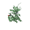
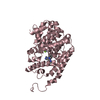
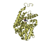
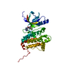
| Title | 3-Sulfinopropionyl-Coenzyme A (3SP-CoA) desulfinase from Advenella mimgardefordensis DPN7T: holo crystal structure with the substrate analog succinyl-CoA | ||||||
 Components Components | ACYL-COA DEHYDROGENASE | ||||||
 Keywords Keywords | OXIDOREDUCTASE | ||||||
| Function / homology |  Function and homology information Function and homology information3-sulfinopropanoyl-CoA desulfinase / acyl-CoA dehydrogenase activity / flavin adenine dinucleotide binding / hydrolase activity Similarity search - Function | ||||||
| Biological species |  ADVENELLA MIMIGARDEFORDENSIS DPN7 (bacteria) ADVENELLA MIMIGARDEFORDENSIS DPN7 (bacteria) | ||||||
| Method |  X-RAY DIFFRACTION / SYNCHROTRON / MOLECULAR REPLACEMENT / Resolution: 2.3 Å X-RAY DIFFRACTION / SYNCHROTRON / MOLECULAR REPLACEMENT / Resolution: 2.3 Å | ||||||
 Authors Authors | Cianci, M. / Schuermann, M. / Meijers, R. / Schneider, T.R. / Steinbuechel, A. | ||||||
 Citation Citation | Journal: Acta Crystallogr.,Sect.D / Year: 2015 Title: 3-Sulfinopropionyl-Coenzyme a (3Sp-Coa) Desulfinase from Advenella Mimigardefordensis Dpn7(T): Crystal Structure and Function of a Desulfinase with an Acyl-Coa Dehydrogenase Fold. Authors: Schurmann, M. / Meijers, R. / Schneider, T.R. / Steinbuchel, A. / Cianci, M. | ||||||
| History |
|
- Structure visualization
| Structure viewer | Molecule: MolmilJmol/JSmol |
|---|
- Downloads & links
-Download
| PDBx/mmCIF format | 5ahs.cif.gz | 491.6 KB | Display | PDBx/mmCIF format |
|---|---|---|---|---|
| PDB format | pdb5ahs.ent.gz | 406.1 KB | Display | PDB format |
| PDBx/mmJSON format | 5ahs.json.gz | Tree view | PDBx/mmJSON format | |
| Others |  Other downloads Other downloads |
-Validation report
| Arichive directory | https://data.pdbj.org/pub/pdb/validation_reports/ah/5ahsftp://data.pdbj.org/pub/pdb/validation_reports/ah/5ahs | HTTPS FTP |
|---|
-Related structure data
| Related structure data |  5af7SC S: Starting model for refinement C: citing same article ( |
|---|---|
| Similar structure data |


-Links
 PDBj
PDBj


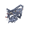
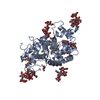
- Assembly
| Deposited unit | 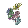
| ||||||||
|---|---|---|---|---|---|---|---|---|---|
| 1 | 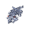
| ||||||||
| 2 | 
| ||||||||
| 3 | 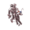
| ||||||||
| 4 | 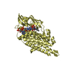
| ||||||||
| 5 | 
| ||||||||
| 6 | 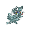
| ||||||||
| Unit cell |
|
-Components
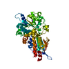
-Protein , 1 types, 6 molecules ABCDEF
| #1: Protein | Mass: 43581.746 Da / Num. of mol.: 6 Source method: isolated from a genetically manipulated source Source: (gene. exp.) ADVENELLA MIMIGARDEFORDENSIS DPN7 (bacteria)Plasmid: PET23A / Production host: References: UniProt: K4L7X3, 3-sulfinopropanoyl-CoA desulfinase |
|---|
-Non-polymers , 5 types, 1475 molecules 


| #2: Chemical | ChemComp-FAD /  Mass: 785.550 Da / Num. of mol.: 6 / Source method: obtained synthetically / Formula: C27H33N9O15P2 / Comment: FAD*YM Mass: 785.550 Da / Num. of mol.: 6 / Source method: obtained synthetically / Formula: C27H33N9O15P2 / Comment: FAD*YM#3: Chemical | ChemComp-COA /  Mass: 767.534 Da / Num. of mol.: 5 / Source method: obtained synthetically / Formula: C21H36N7O16P3S Mass: 767.534 Da / Num. of mol.: 5 / Source method: obtained synthetically / Formula: C21H36N7O16P3S#4: Chemical | ChemComp-SIN / |  Mass: 118.088 Da / Num. of mol.: 1 / Source method: obtained synthetically / Formula: C4H6O4 Mass: 118.088 Da / Num. of mol.: 1 / Source method: obtained synthetically / Formula: C4H6O4#5: Chemical | ChemComp-SO4 / |  Mass: 96.063 Da / Num. of mol.: 1 / Source method: obtained synthetically / Formula: SO4 Mass: 96.063 Da / Num. of mol.: 1 / Source method: obtained synthetically / Formula: SO4#6: Water | ChemComp-HOH / | Mass: 18.015 Da / Num. of mol.: 1462 / Source method: isolated from a natural source / Formula: H2O |
|---|
-Experimental details
-Experiment
| Experiment | Method: X-RAY DIFFRACTION / Number of used crystals: 1 |
|---|
- Sample preparation
| Crystal | Density Matthews: 2.7 Å3/Da / Density % sol: 55.8 % / Description: NONE |
|---|---|
| Crystal grow | Method: vapor diffusion, hanging drop / pH: 7.4 Details: CRYSTALS USED FOR X-RAY DIFFRACTION STUDIES WERE GROWN IN 24-WELL LINBRO PLATES (HAMPTON RESEARCH) BY VAPOUR DIFFUSION IN A HANGING DROP CONFIGURATION FROM 0.1 M BIS- TRIS PH 6.5 AND 5- 20 % ...Details: CRYSTALS USED FOR X-RAY DIFFRACTION STUDIES WERE GROWN IN 24-WELL LINBRO PLATES (HAMPTON RESEARCH) BY VAPOUR DIFFUSION IN A HANGING DROP CONFIGURATION FROM 0.1 M BIS- TRIS PH 6.5 AND 5- 20 % PEG 3350 AT A PROTEIN CONCENTRATION OF 10 MG/ML, BY MIXING 2 UL OF THE PROTEIN SOLUTION WITH 2 UL MOTHER LIQUOR |
-Data collection
| Diffraction | Mean temperature: 100 K |
|---|---|
| Diffraction source | Source: SYNCHROTRON / Site: PETRA III, EMBL c/o DESY  / Beamline: P13 (MX1) / Wavelength: 0.968 / Beamline: P13 (MX1) / Wavelength: 0.968 |
| Detector | Type: DECTRIS PILATUS 6M / Detector: PIXEL / Date: Jun 13, 2013 / Details: KB MIRRORS |
| Radiation | Monochromator: SI(111) / Protocol: SINGLE WAVELENGTH / Monochromatic (M) / Laue (L): M / Scattering type: x-ray |
| Radiation wavelength | Wavelength: 0.968 Å / Relative weight: 1 |
| Reflection | Resolution: 2.3→121.29 Å / Num. obs: 126487 / % possible obs: 99.9 % / Observed criterion σ(I): 2 / Redundancy: 4.9 % / Rmerge(I) obs: 0.08 / Net I/σ(I): 13.5 |
| Reflection shell | Resolution: 2.3→2.34 Å / Redundancy: 5 % / Rmerge(I) obs: 0.95 / Mean I/σ(I) obs: 2 / % possible all: 99.9 |
- Processing
| Software |
| |||||||||||||||||||||||||||||||||||||||||||||||||||||||||||||||||||||||||||||||||||||||||||||||||||||||||||||||||||||||||||||||||||||||||||||||||||||||||||||||||||||||||||||||||||||||||||||||||||||||||||||||||||||||||
|---|---|---|---|---|---|---|---|---|---|---|---|---|---|---|---|---|---|---|---|---|---|---|---|---|---|---|---|---|---|---|---|---|---|---|---|---|---|---|---|---|---|---|---|---|---|---|---|---|---|---|---|---|---|---|---|---|---|---|---|---|---|---|---|---|---|---|---|---|---|---|---|---|---|---|---|---|---|---|---|---|---|---|---|---|---|---|---|---|---|---|---|---|---|---|---|---|---|---|---|---|---|---|---|---|---|---|---|---|---|---|---|---|---|---|---|---|---|---|---|---|---|---|---|---|---|---|---|---|---|---|---|---|---|---|---|---|---|---|---|---|---|---|---|---|---|---|---|---|---|---|---|---|---|---|---|---|---|---|---|---|---|---|---|---|---|---|---|---|---|---|---|---|---|---|---|---|---|---|---|---|---|---|---|---|---|---|---|---|---|---|---|---|---|---|---|---|---|---|---|---|---|---|---|---|---|---|---|---|---|---|---|---|---|---|---|---|---|---|
| Refinement | Method to determine structure: MOLECULAR REPLACEMENT Starting model: PDB ENTRY 5AF7 Resolution: 2.3→116.746 Å / SU ML: 0.31 / σ(F): 1.91 / Phase error: 27.79 / Stereochemistry target values: ML
| |||||||||||||||||||||||||||||||||||||||||||||||||||||||||||||||||||||||||||||||||||||||||||||||||||||||||||||||||||||||||||||||||||||||||||||||||||||||||||||||||||||||||||||||||||||||||||||||||||||||||||||||||||||||||
| Solvent computation | Shrinkage radii: 0.9 Å / VDW probe radii: 1.11 Å / Solvent model: FLAT BULK SOLVENT MODEL | |||||||||||||||||||||||||||||||||||||||||||||||||||||||||||||||||||||||||||||||||||||||||||||||||||||||||||||||||||||||||||||||||||||||||||||||||||||||||||||||||||||||||||||||||||||||||||||||||||||||||||||||||||||||||
| Refinement step | Cycle: LAST / Resolution: 2.3→116.746 Å
| |||||||||||||||||||||||||||||||||||||||||||||||||||||||||||||||||||||||||||||||||||||||||||||||||||||||||||||||||||||||||||||||||||||||||||||||||||||||||||||||||||||||||||||||||||||||||||||||||||||||||||||||||||||||||
| Refine LS restraints |
| |||||||||||||||||||||||||||||||||||||||||||||||||||||||||||||||||||||||||||||||||||||||||||||||||||||||||||||||||||||||||||||||||||||||||||||||||||||||||||||||||||||||||||||||||||||||||||||||||||||||||||||||||||||||||
| LS refinement shell |
|
