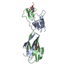
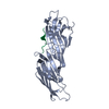
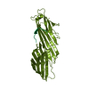
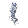
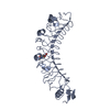
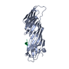
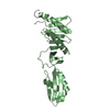
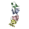
[English] 日本語
Yorodumi
- PDB-5e6v: Re-refinement of the Crystal Structure of the Plexin-Semaphorin-I... -

+
Open data


ID or keywords:

Loading...

-
Basic information

Entry
Database: PDB / ID: 5e6v
TitleRe-refinement of the Crystal Structure of the Plexin-Semaphorin-Integrin Domain/Hybrid Domain/I-EGF1 Segment from the Human Integrin b2 Subunit
ComponentsIntegrin beta-2
KeywordsCELL ADHESION / lymphocyte function-associated antigen-1 / LFA-1
Function / homology
Function and homology information


integrin alphaX-beta2 complex / cellular extravasation / positive regulation of neutrophil degranulation / integrin alphaM-beta2 complex / integrin alphaL-beta2 complex / ICAM-3 receptor activity / cell-cell adhesion via plasma-membrane adhesion molecules / complement component C3b binding / negative regulation of dopamine metabolic process / Toll Like Receptor 4 (TLR4) Cascade ...integrin alphaX-beta2 complex / cellular extravasation / positive regulation of neutrophil degranulation / integrin alphaM-beta2 complex / integrin alphaL-beta2 complex / ICAM-3 receptor activity / cell-cell adhesion via plasma-membrane adhesion molecules / complement component C3b binding / negative regulation of dopamine metabolic process / Toll Like Receptor 4 (TLR4) Cascade / neutrophil migration / leukocyte migration involved in inflammatory response / positive regulation of leukocyte adhesion to vascular endothelial cell / integrin complex / heterotypic cell-cell adhesion / regulation of peptidyl-tyrosine phosphorylation / leukocyte cell-cell adhesion / phagocytosis, engulfment / cell adhesion mediated by integrin / receptor clustering / endodermal cell differentiation / amyloid-beta clearance / tertiary granule membrane / cellular response to low-density lipoprotein particle stimulus / ficolin-1-rich granule membrane / plasma membrane raft / positive regulation of protein targeting to membrane / Integrin cell surface interactions / endothelial cell migration / specific granule membrane / cell adhesion molecule binding / heat shock protein binding / neutrophil chemotaxis / receptor-mediated endocytosis / positive regulation of superoxide anion generation / cell-matrix adhesion / integrin-mediated signaling pathway / Cell surface interactions at the vascular wall / microglial cell activation / cell-cell adhesion / receptor internalization / positive regulation of angiogenesis / Immunoregulatory interactions between a Lymphoid and a non-Lymphoid cell / positive regulation of nitric oxide biosynthetic process / integrin binding / cell-cell signaling / extracellular vesicle / regulation of cell shape / amyloid-beta binding / Interleukin-4 and Interleukin-13 signaling / receptor complex / cell adhesion / inflammatory response / external side of plasma membrane / focal adhesion / apoptotic process / Neutrophil degranulation / protein kinase binding / cell surface / extracellular exosome / metal ion binding / membrane / plasma membrane
Similarity search - Function
Integrin beta-2 subunit / Integrin beta subunit, cytoplasmic domain / Integrin beta cytoplasmic domain / Integrin_b_cyt / Integrin beta tail domain / Integrin EGF domain / Integrin beta subunit, tail / Integrin beta tail domain superfamily / Integrin_B_tail / Integrins beta chain EGF (I-EGF) domain profile. ...Integrin beta-2 subunit / Integrin beta subunit, cytoplasmic domain / Integrin beta cytoplasmic domain / Integrin_b_cyt / Integrin beta tail domain / Integrin EGF domain / Integrin beta subunit, tail / Integrin beta tail domain superfamily / Integrin_B_tail / Integrins beta chain EGF (I-EGF) domain profile. / Integrin beta subunit, VWA domain / Integrin beta subunit / Integrin beta N-terminal / Integrin beta chain VWA domain / Integrin plexin domain / Integrins beta chain EGF (I-EGF) domain signature. / Integrin beta subunits (N-terminal portion of extracellular region) / Integrin domain superfamily / PSI domain / domain found in Plexins, Semaphorins and Integrins / von Willebrand factor A-like domain superfamily / EGF-like domain signature 1. / EGF-like domain signature 2.
Similarity search - Domain/homology
Biological speciesHomo sapiens (human)
MethodX-RAY DIFFRACTION / SAD / Resolution: 1.8 Å
AuthorsSen, M. / Springer, T.A.
Funding support United States, 1items
OrganizationGrant numberCountry
National Institutes of Health/National Cancer Institute (NIH/NCI)NCI CA031798 United States
CitationJournal: J. Biol. Chem. / Year: 2005
Title: The crystal structure of the plexin-semaphorin-integrin domain/hybrid domain/I-EGF1 segment from the human integrin beta2 subunit at 1.8-A resolution.
Authors: Shi, M. / Sundramurthy, K. / Liu, B. / Tan, S.M. / Law, S.K. / Lescar, J.
History
DepositionOct 10, 2015Deposition site: RCSB / Processing site: RCSB
Revision 1.0Mar 2, 2016Provider: repository / Type: Initial release
Revision 1.1Mar 16, 2016Group: Database references
Revision 1.2May 11, 2016Group: Database references
Revision 1.3Mar 15, 2017Group: Experimental preparation / Other
Revision 1.4Sep 27, 2017Group: Author supporting evidence / Category: pdbx_audit_support / Item: _pdbx_audit_support.funding_organization
Revision 2.0Dec 4, 2019Group: Author supporting evidence / Polymer sequence / Category: entity_poly / pdbx_audit_support
Item: _entity_poly.pdbx_seq_one_letter_code_can / _pdbx_audit_support.funding_organization
Revision 3.0Jul 29, 2020Group: Atomic model / Data collection ...Atomic model / Data collection / Derived calculations / Structure summary
Category: atom_site / chem_comp ...atom_site / chem_comp / diffrn_radiation_wavelength / entity / pdbx_branch_scheme / pdbx_chem_comp_identifier / pdbx_entity_branch / pdbx_entity_branch_descriptor / pdbx_entity_branch_link / pdbx_entity_branch_list / pdbx_entity_nonpoly / pdbx_nonpoly_scheme / pdbx_struct_assembly_gen / struct_asym / struct_conn / struct_site / struct_site_gen
Item: _atom_site.B_iso_or_equiv / _atom_site.Cartn_x ..._atom_site.B_iso_or_equiv / _atom_site.Cartn_x / _atom_site.Cartn_y / _atom_site.Cartn_z / _atom_site.auth_asym_id / _atom_site.auth_seq_id / _atom_site.label_asym_id / _atom_site.label_entity_id / _chem_comp.name / _chem_comp.type / _diffrn_radiation_wavelength.wavelength / _entity.formula_weight / _entity.pdbx_description / _entity.pdbx_number_of_molecules / _entity.type / _pdbx_struct_assembly_gen.asym_id_list / _struct_conn.pdbx_role / _struct_conn.ptnr1_auth_asym_id / _struct_conn.ptnr1_auth_seq_id / _struct_conn.ptnr1_label_asym_id / _struct_conn.ptnr1_label_atom_id / _struct_conn.ptnr2_auth_asym_id / _struct_conn.ptnr2_auth_seq_id / _struct_conn.ptnr2_label_asym_id / _struct_conn.ptnr2_label_atom_id
Description: Carbohydrate remediation / Provider: repository / Type: Remediation
Revision 3.1Nov 6, 2024Group: Data collection / Database references ...Data collection / Database references / Derived calculations / Structure summary
Category: chem_comp / chem_comp_atom ...chem_comp / chem_comp_atom / chem_comp_bond / database_2 / pdbx_entry_details / pdbx_modification_feature / struct_conn
Item: _chem_comp.pdbx_synonyms / _database_2.pdbx_DOI ..._chem_comp.pdbx_synonyms / _database_2.pdbx_DOI / _database_2.pdbx_database_accession / _struct_conn.pdbx_leaving_atom_flag
Remark 0This entry 5E6V reflects an alternative modeling of the structural data in r1YUKSF original data ...This entry 5E6V reflects an alternative modeling of the structural data in r1YUKSF original data determined by Authors: Shi, M., Sundramurthy, K., Liu, B., Tan, S.M., Law, S.K., Lescar, J.

-
Structure visualization

Structure viewerMolecule:
MolmilJmol/JSmol

Downloads & links

-
Assembly

Deposited unit
A: Integrin beta-2
hetero molecules


Theoretical massNumber of molelcules
Total (without water)25,9683
Polymers24,9731
Non-polymers9952
Water4,234235
1


  • Idetical with deposited unit
  • defined by author&software
TypeNameSymmetry operationNumber
identity operation1_555x,y,z1
MethodPISA
Unit cell
Length a, b, c (Å)58.600, 31.818, 74.950
Angle α, β, γ (deg.)90.00, 91.20, 90.00
Int Tables number4
Space group name H-MP1211

-
Components

#1: Protein Integrin beta-2 / Cell surface adhesion glycoproteins LFA-1/CR3/p150 / 95 subunit beta / Complement receptor C3 subunit beta


Mass: 24973.260 Da / Num. of mol.: 1 / Fragment: UNP residues 23-125, 365-482
Source method: isolated from a genetically manipulated source
Source: (gene. exp.) Homo sapiens (human) / Gene: ITGB2, CD18, MFI7 / Cell line (production host): HEK / Production host: Homo sapiens (human) / References: UniProt: P05107
#2: Polysaccharide 2-acetamido-2-deoxy-beta-D-glucopyranose-(1-4)-2-acetamido-2-deoxy-beta-D-glucopyranose


Type: oligosaccharide / Mass: 424.401 Da / Num. of mol.: 1
Source method: isolated from a genetically manipulated source
DescriptorTypeProgram
DGlcpNAcb1-4DGlcpNAcb1-Glycam Condensed SequenceGMML 1.0
WURCS=2.0/1,2,1/[a2122h-1b_1-5_2*NCC/3=O]/1-1/a4-b1WURCSPDB2Glycan 1.1.0
[]{[(4+1)][b-D-GlcpNAc]{[(4+1)][b-D-GlcpNAc]{}}}LINUCSPDB-CARE
#3: Polysaccharide 2-acetamido-2-deoxy-beta-D-glucopyranose-(1-4)-[beta-L-fucopyranose-(1-6)]2-acetamido-2-deoxy-beta- ...2-acetamido-2-deoxy-beta-D-glucopyranose-(1-4)-[beta-L-fucopyranose-(1-6)]2-acetamido-2-deoxy-beta-D-glucopyranose


Type: oligosaccharide / Mass: 570.542 Da / Num. of mol.: 1
Source method: isolated from a genetically manipulated source
DescriptorTypeProgram
DGlcpNAcb1-4[LFucpb1-6]DGlcpNAcb1-Glycam Condensed SequenceGMML 1.0
WURCS=2.0/2,3,2/[a2122h-1b_1-5_2*NCC/3=O][a1221m-1b_1-5]/1-1-2/a4-b1_a6-c1WURCSPDB2Glycan 1.1.0
[]{[(4+1)][b-D-GlcpNAc]{[(4+1)][b-D-GlcpNAc]{}[(6+1)][b-L-Fucp]{}}}LINUCSPDB-CARE
#4: Water ChemComp-HOH / water


Mass: 18.015 Da / Num. of mol.: 235 / Source method: isolated from a natural source / Formula: H2O
Has protein modificationY

-
Experimental details

-
Experiment

ExperimentMethod: X-RAY DIFFRACTION

-
Sample preparation

CrystalDensity Matthews: 2.8 Å3/Da / Density % sol: 56.03 % / Description: AUTHOR USED THE SF DATA FROM ENTRY 1YUK

-
Data collection

RadiationProtocol: MAD / Monochromatic (M) / Laue (L): M / Scattering type: x-ray
Radiation wavelengthRelative weight: 1

-
Processing

Software
NameVersionClassification
PHENIXdev_1839refinement
SOLVEphasing
RefinementMethod to determine structure: SAD / Resolution: 1.8→27.479 Å / SU ML: 0.2 / Cross valid method: FREE R-VALUE / σ(F): 1.35 / Phase error: 22.86 / Stereochemistry target values: ML
RfactorNum. reflection% reflectionSelection details
Rfree0.2293 1322 5.09 %Random selection
Rwork0.1768 ---
obs0.1795 24802 99.28 %-
Solvent computationShrinkage radii: 0.9 Å / VDW probe radii: 1.11 Å / Solvent model: FLAT BULK SOLVENT MODEL
Refinement stepCycle: LAST / Resolution: 1.8→27.479 Å
ProteinNucleic acidLigandSolventTotal
Num. atoms1722 0 66 235 2023
Refine LS restraints
Refine-IDTypeDev idealNumber
X-RAY DIFFRACTIONf_bond_d0.0091840
X-RAY DIFFRACTIONf_angle_d1.2132506
X-RAY DIFFRACTIONf_dihedral_angle_d12.294701
X-RAY DIFFRACTIONf_chiral_restr0.049288
X-RAY DIFFRACTIONf_plane_restr0.005327
LS refinement shell
Resolution (Å)Rfactor RfreeNum. reflection RfreeRfactor RworkNum. reflection RworkRefine-ID% reflection obs (%)
1.8-1.8720.27941240.25132634X-RAY DIFFRACTION96
1.872-1.95720.27251300.21272727X-RAY DIFFRACTION100
1.9572-2.06040.25261440.19332717X-RAY DIFFRACTION100
2.0604-2.18940.24181640.18522713X-RAY DIFFRACTION100
2.1894-2.35840.21371460.17182750X-RAY DIFFRACTION100
2.3584-2.59560.23361330.18572760X-RAY DIFFRACTION100
2.5956-2.97070.21871650.17762727X-RAY DIFFRACTION100
2.9707-3.74130.22611510.16182780X-RAY DIFFRACTION100
3.7413-27.4820.22331650.16932830X-RAY DIFFRACTION99
Refinement TLS params.

Method: refined / Refine-ID: X-RAY DIFFRACTION

IDL112)L122)L132)L222)L232)L332)S11 (Å °)S12 (Å °)S13 (Å °)S21 (Å °)S22 (Å °)S23 (Å °)S31 (Å °)S32 (Å °)S33 (Å °)T112)T122)T132)T222)T232)T332)Origin x (Å)Origin y (Å)Origin z (Å)
10.9497-0.3717-0.1631.112-0.06711.27690.01570.0542-0.2031-0.0810.157-0.11820.26610.2669-00.16240.0133-0.02350.1633-0.03510.257322.53456.9731-1.3187
21.5702-0.1857-0.29230.2368-0.10621.38720.72730.03161.21610.1461-0.2289-0.0665-1.005-0.4175-0.00350.31840.09140.08530.1865-0.0950.3706-2.161616.413516.1344
30.5807-0.07990.47731.238-0.15990.3292-0.1321-0.26670.28150.4025-0.0079-0.10820.0444-0.1693-0.01180.28910.0168-0.01260.2459-0.04270.1921-2.6559.772420.3146
40.9691-0.2237-0.32610.69850.0950.5914-0.1605-0.4699-0.29240.2230.0799-0.27660.27020.0972-0.27930.28780.0373-0.07520.3510.04340.1199-2.6812.510925.3735
50.19380.5057-0.30190.0236-0.17960.8357-0.01310.17150.16880.02630.129-0.0567-0.0427-0.2190.00110.20010.01740.03460.16880.01870.22560.871711.3474.9565
60.3203-0.07180.23480.65790.14250.9789-0.04110.2844-0.3788-0.1931-0.0162-0.31450.3710.42450.0780.24120.01480.06280.3458-0.07960.247422.066214.2841-19.5924
Refinement TLS group
IDRefine-IDRefine TLS-IDSelection details
1X-RAY DIFFRACTION1chain 'A' and (resid 2 through 55 )
2X-RAY DIFFRACTION2chain 'A' and (resid 56 through 79 )
3X-RAY DIFFRACTION3chain 'A' and (resid 80 through 343 )
4X-RAY DIFFRACTION4chain 'A' and (resid 344 through 395 )
5X-RAY DIFFRACTION5chain 'A' and (resid 396 through 435 )
6X-RAY DIFFRACTION6chain 'A' and (resid 436 through 463 )

+
About Yorodumi

-
News

-
Feb 9, 2022. New format data for meta-information of EMDB entries

New format data for meta-information of EMDB entries

  • Version 3 of the EMDB header file is now the official format.
  • The previous official version 1.9 will be removed from the archive.

Related info.:EMDB header

External links:wwPDB to switch to version 3 of the EMDB data model

-
Aug 12, 2020. Covid-19 info

Covid-19 info

URL: https://pdbj.org/emnavi/covid19.php

New page: Covid-19 featured information page in EM Navigator.

Related info.:Covid-19 info / Mar 5, 2020. Novel coronavirus structure data

+
Mar 5, 2020. Novel coronavirus structure data

Novel coronavirus structure data

Related info.:Yorodumi Speices / Aug 12, 2020. Covid-19 info

External links:COVID-19 featured content - PDBj / Molecule of the Month (242):Coronavirus Proteases

+
Jan 31, 2019. EMDB accession codes are about to change! (news from PDBe EMDB page)

EMDB accession codes are about to change! (news from PDBe EMDB page)

  • The allocation of 4 digits for EMDB accession codes will soon come to an end. Whilst these codes will remain in use, new EMDB accession codes will include an additional digit and will expand incrementally as the available range of codes is exhausted. The current 4-digit format prefixed with “EMD-” (i.e. EMD-XXXX) will advance to a 5-digit format (i.e. EMD-XXXXX), and so on. It is currently estimated that the 4-digit codes will be depleted around Spring 2019, at which point the 5-digit format will come into force.
  • The EM Navigator/Yorodumi systems omit the EMD- prefix.

Related info.:Q: What is EMD? / ID/Accession-code notation in Yorodumi/EM Navigator

External links:EMDB Accession Codes are Changing Soon! / Contact to PDBj

+
Jul 12, 2017. Major update of PDB

Major update of PDB

  • wwPDB released updated PDB data conforming to the new PDBx/mmCIF dictionary.
  • This is a major update changing the version number from 4 to 5, and with Remediation, in which all the entries are updated.
  • In this update, many items about electron microscopy experimental information are reorganized (e.g. em_software).
  • Now, EM Navigator and Yorodumi are based on the updated data.

External links:wwPDB Remediation / Enriched Model Files Conforming to OneDep Data Standards Now Available in the PDB FTP Archive

-
Yorodumi

Thousand views of thousand structures

  • Yorodumi is a browser for structure data from EMDB, PDB, SASBDB, etc.
  • This page is also the successor to EM Navigator detail page, and also detail information page/front-end page for Omokage search.
  • The word "yorodu" (or yorozu) is an old Japanese word meaning "ten thousand". "mi" (miru) is to see.

Related info.:EMDB / PDB / SASBDB / Comparison of 3 databanks / Yorodumi Search / Aug 31, 2016. New EM Navigator & Yorodumi / Yorodumi Papers / Jmol/JSmol / Function and homology information / Changes in new EM Navigator and Yorodumi

Read more