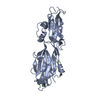
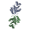
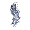
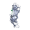
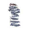
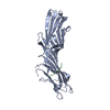
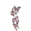
- PDB-4l3r: Crystal structure of a DUF4847 family protein (BACEGG_01241) from... -
+
Open data
ID or keywords:
Loading...
-
Basic information
Entry
Database: PDB / ID: 4l3r
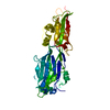
Title
Crystal structure of a DUF4847 family protein (BACEGG_01241) from Bacteroides eggerthii DSM 20697 at 2.23 A resolution
Components
Uncharacterized protein
Keywords
STRUCTURAL GENOMICS / UNKNOWN FUNCTION / PF16139 family / DUF4847 / Joint Center for Structural Genomics / JCSG / Protein Structure Initiative / PSI-BIOLOGY
Mass: 18.015 Da / Num. of mol.: 151 / Source method: isolated from a natural source / Formula: H2O
Has protein modification
Y
Sequence details
THIS CONSTRUCT WAS EXPRESSED WITH A PURIFICATION TAG MGSDKIHHHHHHENLYFQG. THE TAG WAS REMOVED WITH ...THIS CONSTRUCT WAS EXPRESSED WITH A PURIFICATION TAG MGSDKIHHHHHHENLYFQG. THE TAG WAS REMOVED WITH TEV PROTEASE LEAVING ONLY A GLYCINE (0) FOLLOWED BY RESIDUES 28-172 OF THE TARGET SEQUENCE.
-
Experimental details
-
Experiment
Experiment
Method: X-RAY DIFFRACTION / Number of used crystals: 1
-
Sample preparation
Crystal
Density Matthews: 2.67 Å3/Da / Density % sol: 53.96 %
Crystal grow
Temperature: 293 K / Method: vapor diffusion, sitting drop Details: 0.1M citric acid pH 5, 20% polyethylene glycol 6000, NANODROP, VAPOR DIFFUSION, SITTING DROP, temperature 293K
Type: DECTRIS PILATUS 6M / Detector: PIXEL / Date: Mar 21, 2013 Details: Flat mirror (vertical focusing); single crystal Si(111) bent monochromator (horizontal focusing)
Radiation
Monochromator: single crystal Si(111) bent / Protocol: MAD / Monochromatic (M) / Laue (L): M / Scattering type: x-ray
Radiation wavelength
ID
Wavelength (Å)
Relative weight
1
0.91837
1
2
0.97949
1
3
0.97886
1
Reflection
Resolution: 2.23→29.383 Å / Num. obs: 17344 / % possible obs: 95 % / Observed criterion σ(I): -3 / Biso Wilson estimate: 61.075 Å2 / Rmerge(I) obs: 0.028 / Net I/σ(I): 15.48
Reflection shell
Resolution (Å)
Rmerge(I) obs
Mean I/σ(I) obs
Num. measured obs
Num. unique obs
Diffraction-ID
% possible all
2.23-2.31
0.532
1.4
5971
3103
1
93.3
2.31-2.4
0.381
2
5609
2967
1
92.5
2.4-2.51
0.261
2.7
6581
3280
1
98.3
2.51-2.64
0.169
4.2
6296
3162
1
97.2
2.64-2.81
0.106
6.6
6604
3299
1
96.9
2.81-3.02
0.063
10.8
5729
2989
1
93.5
3.02-3.33
0.038
17.9
6466
3252
1
95.6
3.33-3.81
0.023
29.4
6372
3174
1
96
3.81-4.78
0.018
37.2
5761
3003
1
92
4.78-29.383
0.014
42.9
6450
3198
1
94.6
-
Phasing
Phasing
Method: MAD
-
Processing
Software
Name
Version
Classification
NB
MolProbity
3beta29
modelbuilding
PDB_EXTRACT
3.1
dataextraction
SHELX
phasing
SHARP
phasing
XSCALE
July4, 2012
datascaling
BUSTER-TNT
2.10.0
refinement
XDS
datareduction
SHELXD
phasing
BUSTER
2.10.0
refinement
Refinement
Method to determine structure: MAD / Resolution: 2.23→29.383 Å / Cor.coef. Fo:Fc: 0.9528 / Cor.coef. Fo:Fc free: 0.9365 / Occupancy max: 1 / Occupancy min: 0.22 / Cross valid method: THROUGHOUT / σ(F): 0 Details: 1. ZERO OCCUPANCY HYDROGENS WERE INCLUDED DURING REFINEMENT TO IMPROVE THE ANTI-BUMPING RESTRAINTS. 2. ATOM RECORDS CONTAIN SUM OF TLS AND RESIDUAL B FACTORS. 3. ANISOU RECORDS CONTAIN SUM ...Details: 1. ZERO OCCUPANCY HYDROGENS WERE INCLUDED DURING REFINEMENT TO IMPROVE THE ANTI-BUMPING RESTRAINTS. 2. ATOM RECORDS CONTAIN SUM OF TLS AND RESIDUAL B FACTORS. 3. ANISOU RECORDS CONTAIN SUM OF TLS AND RESIDUAL U FACTORS. 4. WATERS WERE EXCLUDED FROM AUTOMATIC TLS ASSIGNMENT. 5. A MET-INHIBITION PROTOCOL WAS USED FOR SELENOMETHIONINE INCORPORATION DURING PROTEIN EXPRESSION. THE OCCUPANCY OF THE SE ATOMS IN THE MSE RESIDUES WAS REDUCED TO 0.75 FOR THE REDUCED SCATTERING POWER DUE TO PARTIAL S-MET INCORPORATION. 6. MAD PHASE RESTRAINTS WERE USED DURING REFINEMENT. 7. NCS RESTRAINTS WERE APPLIED DURING REFINEMENT USING LSSR (-AUTONCS) IN BUSTER.
In the structure databanks used in Yorodumi, some data are registered as the other names, "COVID-19 virus" and "2019-nCoV". Here are the details of the virus and the list of structure data.
Jan 31, 2019. EMDB accession codes are about to change! (news from PDBe EMDB page)
EMDB accession codes are about to change! (news from PDBe EMDB page)
The allocation of 4 digits for EMDB accession codes will soon come to an end. Whilst these codes will remain in use, new EMDB accession codes will include an additional digit and will expand incrementally as the available range of codes is exhausted. The current 4-digit format prefixed with “EMD-” (i.e. EMD-XXXX) will advance to a 5-digit format (i.e. EMD-XXXXX), and so on. It is currently estimated that the 4-digit codes will be depleted around Spring 2019, at which point the 5-digit format will come into force.
The EM Navigator/Yorodumi systems omit the EMD- prefix.
Related info.:Q: What is EMD? / ID/Accession-code notation in Yorodumi/EM Navigator
Yorodumi is a browser for structure data from EMDB, PDB, SASBDB, etc.
This page is also the successor to EM Navigator detail page, and also detail information page/front-end page for Omokage search.
The word "yorodu" (or yorozu) is an old Japanese word meaning "ten thousand". "mi" (miru) is to see.
Related info.:EMDB / PDB / SASBDB / Comparison of 3 databanks / Yorodumi Search / Aug 31, 2016. New EM Navigator & Yorodumi / Yorodumi Papers / Jmol/JSmol / Function and homology information / Changes in new EM Navigator and Yorodumi
 Movie
Movie Controller

 Yorodumi
 Open data
 Basic information
 Components
 Keywords
 Function and homology information
 Bacteroides eggerthii (bacteria)
 X-RAY DIFFRACTION /
  Authors
 Citation
 Structure visualization
 Downloads & links
 Other downloads
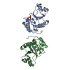


 PDBj
PDBj Assembly
 Mass: 18.015 Da / Num. of mol.: 151 / Source method: isolated from a natural source / Formula: H2O
 Sample preparation
 / Beamline: BL11-1 / Wavelength: 0.91837,0.97949,0.97886
 Processing