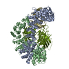
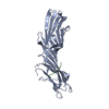
SHEET THE SHEET STRUCTURE OF THIS MOLECULE IS BIFURCATED. IN ORDER TO REPRESENT THIS FEATURE IN ... SHEET THE SHEET STRUCTURE OF THIS MOLECULE IS BIFURCATED. IN ORDER TO REPRESENT THIS FEATURE IN THE SHEET RECORDS BELOW, TWO SHEETS ARE DEFINED.
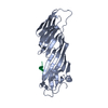
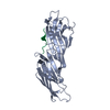
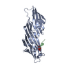
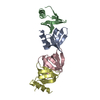
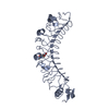
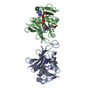
ALTHOUGH CHAIN M IS A DOMAIN OF A LARGER PROTEINWHICH EXISTS IN ITS NATIVE STATE AS A TETRAMER, THISENTRY IS ANNOTATED AS A DIMER AS IT IS IN COMPLEXWITH A SMALL PEPTIDE (CHAIN P).
-
Components
#1: Protein
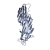
CLATHRINCOATASSEMBLYPROTEINAP50 / CLATHRIN COAT ASSOCIATED PROTEIN AP50 / PLASMA MEMBRANE ADAPTOR AP-2 50 KDA PROTEIN / CLATHRIN ...CLATHRIN COAT ASSOCIATED PROTEIN AP50 / PLASMA MEMBRANE ADAPTOR AP-2 50 KDA PROTEIN / CLATHRIN ASSEMBLY PROTEIN COMPLEX 2 MEDIUM CHAIN / AP-2 MU 2 CHAIN
Mass: 49726.641 Da / Num. of mol.: 1 Source method: isolated from a genetically manipulated source Source: (gene. exp.) RATTUS NORVEGICUS (Norway rat) / Plasmid: PMW172H6 / Production host: ESCHERICHIA COLI (E. coli) / Strain (production host): BL21 / References: UniProt: P84092
#2: Protein/peptide
P2XPURINOCEPTOR4 / ATP RECEPTOR / P2X4 / PURINERGIC RECEPTOR
Mass: 18.015 Da / Num. of mol.: 69 / Source method: isolated from a natural source / Formula: H2O
Compound details
FUNCTION: COMPONENT OF THE ADAPTOR COMPLEXES WHICH LINK CLATHRIN TO RECEPTORS IN COATED VESICLES. ...FUNCTION: COMPONENT OF THE ADAPTOR COMPLEXES WHICH LINK CLATHRIN TO RECEPTORS IN COATED VESICLES. CLATHRIN-ASSOCIATED PROTEIN COMPLEXES ARE BELIEVED TO INTERACT WITH THE CYTOPLASMIC TAILS OF MEMBRANE PROTEINS, LEADING TO THEIR SELECTION AND CONCENTRATION. AP50 IS A SUBUNIT OF THE PLASMA MEMBRANE ADAPTOR. THE COMPLEX BINDS POLYPHOSPHOINOSITIDE-CONTAINING LIPIDS.
-
Experimental details
-
Experiment
Experiment
Method: X-RAY DIFFRACTION / Number of used crystals: 1
-
Sample preparation
Crystal
Density Matthews: 2.8 Å3/Da / Density % sol: 60 % / Description: ISOMORPHOUS CRYSTAL TO 1BXX
Resolution: 2.8→105.41 Å / Cor.coef. Fo:Fc: 0.954 / Cor.coef. Fo:Fc free: 0.915 / SU B: 9.807 / SU ML: 0.184 / Cross valid method: THROUGHOUT / ESU R: 0.305 / ESU R Free: 0.266 / Stereochemistry target values: MAXIMUM LIKELIHOOD / Details: HYDROGENS HAVE BEEN ADDED IN THE RIDING POSITIONS.
Rfactor
Num. reflection
% reflection
Selection details
Rfree
0.251
825
5 %
RANDOM
Rwork
0.192
-
-
-
obs
0.195
15655
99.8 %
-
Solvent computation
Ion probe radii: 0.8 Å / Shrinkage radii: 0.8 Å / VDW probe radii: 1.2 Å / Solvent model: BABINET MODEL WITH MASK
 Movie
Movie Controller

 Yorodumi
 Open data
 Basic information
 Components
 Keywords
 Function and homology information
 X-RAY DIFFRACTION /
  Authors
 Citation
 Structure visualization
 Downloads & links
 Other downloads
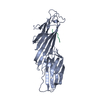


 PDBj
PDBj


 Assembly
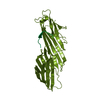

 Mass: 18.015 Da / Num. of mol.: 69 / Source method: isolated from a natural source / Formula: H2O
 Sample preparation
 / Beamline: PX14.2 / Wavelength: 1
  Processing