 Movie
Movie Controller

[English] 日本語
 Yorodumi
- PDB-5dnb: STRUCTURE OF THE B-DNA DECAMER C-C-A-A-C-G-T-T-G-G AND COMPARISON... -
+ Open data

- Basic information
| Entry | Database: PDB / ID: 5dnb | |||||||||||||||||||||
|---|---|---|---|---|---|---|---|---|---|---|---|---|---|---|---|---|---|---|---|---|---|---|
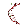
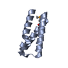
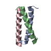
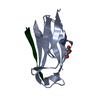
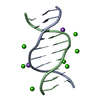
| Title | STRUCTURE OF THE B-DNA DECAMER C-C-A-A-C-G-T-T-G-G AND COMPARISON WITH ISOMORPHOUS DECAMERS C-C-A-A-G-A-T-T-G-G AND C-C-A-G-G-C-C-T-G-G | |||||||||||||||||||||
 Components Components | DNA (5'-D(* Keywords KeywordsDNA / B-DNA / DOUBLE HELIX | Function / homology | DNA |  Function and homology information Function and homology informationMethod |  X-RAY DIFFRACTION / Resolution: 1.4 Å X-RAY DIFFRACTION / Resolution: 1.4 Å  Authors AuthorsPrive, G.G. / Yanagi, K. / Dickerson, R.E. |  CitationJournal: J.Mol.Biol. / Year: 1991 CitationJournal: J.Mol.Biol. / Year: 1991Title: Structure of the B-DNA decamer C-C-A-A-C-G-T-T-G-G and comparison with isomorphous decamers C-C-A-A-G-A-T-T-G-G and C-C-A-G-G-C-C-T-G-G. Authors: Prive, G.G. / Yanagi, K. / Dickerson, R.E. History |
|
- Structure visualization
| Structure viewer | Molecule: MolmilJmol/JSmol |
|---|
- Downloads & links
-Download
| PDBx/mmCIF format | 5dnb.cif.gz | 18.6 KB | Display | PDBx/mmCIF format |
|---|---|---|---|---|
| PDB format | pdb5dnb.ent.gz | 10.5 KB | Display | PDB format |
| PDBx/mmJSON format | 5dnb.json.gz | Tree view | PDBx/mmJSON format | |
| Others |  Other downloads Other downloads |
-Validation report
| Arichive directory | https://data.pdbj.org/pub/pdb/validation_reports/dn/5dnbftp://data.pdbj.org/pub/pdb/validation_reports/dn/5dnb | HTTPS FTP |
|---|
-Related structure data


-Links
 PDBj
PDBj

- Assembly
| Deposited unit | 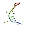
| ||||||||
|---|---|---|---|---|---|---|---|---|---|
| 1 | 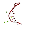
| ||||||||
| Unit cell |
| ||||||||
| Atom site foot note | 1: THESE WATER MOLECULES MAKE A SPLINE OF HYDRATION. / 2: THESE WATER MOLECULES MAKE A SPLINE OF HYDRATION. / 3: THESE WATER MOLECULES MAKE A SPLINE OF HYDRATION. / 4: HOH 63 IS ONLY 1.57 ANGSTROMS FROM THE O4* OF A 8. | ||||||||
| Components on special symmetry positions |
|
-Components
| #1: DNA chain | Mass: 3045.004 Da / Num. of mol.: 1 / Source method: obtained synthetically | ||
|---|---|---|---|
| #2: Chemical | ChemComp-MG /   Mass: 24.305 Da / Num. of mol.: 4 / Source method: obtained synthetically / Formula: Mg Mass: 24.305 Da / Num. of mol.: 4 / Source method: obtained synthetically / Formula: Mg#3: Water | ChemComp-HOH / |  Mass: 18.015 Da / Num. of mol.: 77 / Source method: isolated from a natural source / Formula: H2O Mass: 18.015 Da / Num. of mol.: 77 / Source method: isolated from a natural source / Formula: H2O |
-Experimental details
-Experiment
| Experiment | Method: X-RAY DIFFRACTION |
|---|
- Sample preparation
| Crystal | Density Matthews: 2.05 Å3/Da / Density % sol: 39.91 % | ||||||||||||||||||||||||||||||||||||
|---|---|---|---|---|---|---|---|---|---|---|---|---|---|---|---|---|---|---|---|---|---|---|---|---|---|---|---|---|---|---|---|---|---|---|---|---|---|
| Crystal grow | Method: vapor diffusion / Details: VAPOR DIFFUSION | ||||||||||||||||||||||||||||||||||||
| Components of the solutions |
| ||||||||||||||||||||||||||||||||||||
| Crystal grow | *PLUS Method: vapor diffusion | ||||||||||||||||||||||||||||||||||||
| Components of the solutions | *PLUS
|
-Data collection
| Diffraction | Mean temperature: 273 K |
|---|---|
| Diffraction source | Source: ROTATING ANODE |
| Detector | Type: RIGAKU AFC-5R / Detector: DIFFRACTOMETER |
| Radiation | Protocol: SINGLE WAVELENGTH / Monochromatic (M) / Laue (L): M / Scattering type: x-ray |
| Radiation wavelength | Relative weight: 1 |
| Reflection | Resolution: 1.4→8 Å / Num. obs: 4402 / Observed criterion σ(I): 1 |
- Processing
| Software | Name: NUCLSQ / Classification: refinement | ||||||||||||||||||||||||||||||||||||||||||||||||||||||||||||||||||||||||||||||||||||
|---|---|---|---|---|---|---|---|---|---|---|---|---|---|---|---|---|---|---|---|---|---|---|---|---|---|---|---|---|---|---|---|---|---|---|---|---|---|---|---|---|---|---|---|---|---|---|---|---|---|---|---|---|---|---|---|---|---|---|---|---|---|---|---|---|---|---|---|---|---|---|---|---|---|---|---|---|---|---|---|---|---|---|---|---|---|
| Refinement | Resolution: 1.4→8 Å / σ(F): 1 /
| ||||||||||||||||||||||||||||||||||||||||||||||||||||||||||||||||||||||||||||||||||||
| Refine Biso |
| ||||||||||||||||||||||||||||||||||||||||||||||||||||||||||||||||||||||||||||||||||||
| Refinement step | Cycle: LAST / Resolution: 1.4→8 Å
| ||||||||||||||||||||||||||||||||||||||||||||||||||||||||||||||||||||||||||||||||||||
| Refine LS restraints |
| ||||||||||||||||||||||||||||||||||||||||||||||||||||||||||||||||||||||||||||||||||||
| Refinement | *PLUS Highest resolution: 1.4 Å / Num. reflection all: 4402 / σ(F): 1 / Rfactor all: 0.16 | ||||||||||||||||||||||||||||||||||||||||||||||||||||||||||||||||||||||||||||||||||||
| Solvent computation | *PLUS | ||||||||||||||||||||||||||||||||||||||||||||||||||||||||||||||||||||||||||||||||||||
| Displacement parameters | *PLUS |
