 Movie
Movie Controller


+ Open data

- Basic information
| Entry | Database: PDB / ID: 4r49 | ||||||||||||||||||||
|---|---|---|---|---|---|---|---|---|---|---|---|---|---|---|---|---|---|---|---|---|---|
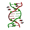
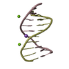
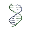
| Title | Racemic crystal structure of a calcium-bound B-DNA duplex | ||||||||||||||||||||
 Components Components | 5'-D(* Keywords KeywordsDNA / racemic DNA / racemates | Function / homology | DNA |  Function and homology information Function and homology informationBiological species | synthetic construct (others) | Method |  X-RAY DIFFRACTION / SYNCHROTRON / MOLECULAR REPLACEMENT / Resolution: 1.28 Å X-RAY DIFFRACTION / SYNCHROTRON / MOLECULAR REPLACEMENT / Resolution: 1.28 Å  Authors AuthorsMandal, P.K. / Collie, G.W. / Kauffmann, B. / Huc, I. |  CitationJournal: Angew.Chem.Int.Ed.Engl. / Year: 2014 CitationJournal: Angew.Chem.Int.Ed.Engl. / Year: 2014Title: Racemic DNA crystallography. Authors: Mandal, P.K. / Collie, G.W. / Kauffmann, B. / Huc, I. History |
|
- Structure visualization
| Structure viewer | Molecule: MolmilJmol/JSmol |
|---|
- Downloads & links
-Download
| PDBx/mmCIF format | 4r49.cif.gz | 40.9 KB | Display | PDBx/mmCIF format |
|---|---|---|---|---|
| PDB format | pdb4r49.ent.gz | 29.1 KB | Display | PDB format |
| PDBx/mmJSON format | 4r49.json.gz | Tree view | PDBx/mmJSON format | |
| Others |  Other downloads Other downloads |
-Validation report
| Arichive directory | https://data.pdbj.org/pub/pdb/validation_reports/r4/4r49ftp://data.pdbj.org/pub/pdb/validation_reports/r4/4r49 | HTTPS FTP |
|---|
-Related structure data
| Related structure data | 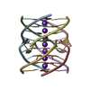 4r44C 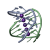 4r45C 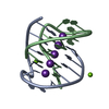 4r47C  4r48C 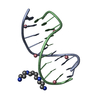 4r4aC 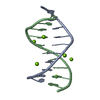 4r4dC 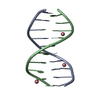 3r86S C: citing same article ( S: Starting model for refinement |
|---|---|
| Similar structure data |


-Links
 PDBj
PDBj

- Assembly
| Deposited unit | 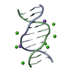
| ||||||||
|---|---|---|---|---|---|---|---|---|---|
| 1 |
| ||||||||
| Unit cell |
| ||||||||
| Components on special symmetry positions |
|
-Components
| #1: DNA chain | Mass: 3045.992 Da / Num. of mol.: 2 / Source method: obtained synthetically / Details: B-DNA Duplex / Source: (synth.) synthetic construct (others) #2: Chemical | ChemComp-CA /   Mass: 40.078 Da / Num. of mol.: 7 / Source method: obtained synthetically / Formula: Ca Mass: 40.078 Da / Num. of mol.: 7 / Source method: obtained synthetically / Formula: Ca#3: Chemical |   Mass: 22.990 Da / Num. of mol.: 2 / Source method: obtained synthetically / Formula: Na Mass: 22.990 Da / Num. of mol.: 2 / Source method: obtained synthetically / Formula: Na#4: Water | ChemComp-HOH / |  Mass: 18.015 Da / Num. of mol.: 167 / Source method: isolated from a natural source / Formula: H2O Mass: 18.015 Da / Num. of mol.: 167 / Source method: isolated from a natural source / Formula: H2O |
|---|
-Experimental details
-Experiment
| Experiment | Method: X-RAY DIFFRACTION / Number of used crystals: 1 |
|---|
- Sample preparation
| Crystal | Density Matthews: 2.07 Å3/Da / Density % sol: 40.51 % |
|---|---|
| Crystal grow | Temperature: 293 K / Method: vapor diffusion, hanging drop / pH: 7 Details: 1 mM DNA, 50 mM sodium cacodylate, 2 M calcium chloride, 1 mM spermine, 40% v/v MPD, pH 7.0, VAPOR DIFFUSION, HANGING DROP, temperature 293K |
-Data collection
| Diffraction | Mean temperature: 100 K |
|---|---|
| Diffraction source | Source: SYNCHROTRON / Site: ESRF  / Beamline: ID23-2 / Wavelength: 0.873 Å / Beamline: ID23-2 / Wavelength: 0.873 Å |
| Detector | Type: DECTRIS PILATUS 2M-F / Detector: PIXEL / Date: Jun 14, 2014 |
| Radiation | Monochromator: Si(111) / Protocol: SINGLE WAVELENGTH / Monochromatic (M) / Laue (L): M / Scattering type: x-ray |
| Radiation wavelength | Wavelength: 0.873 Å / Relative weight: 1 |
| Reflection | Resolution: 1.28→34.25 Å / Num. all: 25176 / Num. obs: 23975 / % possible obs: 95.23 % / Observed criterion σ(F): 2 / Observed criterion σ(I): 2 / Redundancy: 1.9 % / Biso Wilson estimate: 47.43 Å2 / Rmerge(I) obs: 0.0372 / Net I/σ(I): 9.69 |
| Reflection shell | Resolution: 1.28→1.32 Å / Redundancy: 1.8 % / Rmerge(I) obs: 0.2656 / Mean I/σ(I) obs: 2.39 / % possible all: 91.68 |
- Processing
| Software |
| ||||||||||||||||||||||||||||||||||||||||
|---|---|---|---|---|---|---|---|---|---|---|---|---|---|---|---|---|---|---|---|---|---|---|---|---|---|---|---|---|---|---|---|---|---|---|---|---|---|---|---|---|---|
| Refinement | Method to determine structure: MOLECULAR REPLACEMENT Starting model: PDB ENTRY 3R86 Resolution: 1.28→34.25 Å / Cor.coef. Fo:Fc: 0.961 / Cor.coef. Fo:Fc free: 0.943 / SU B: 1.437 / SU ML: 0.027 / Cross valid method: THROUGHOUT / ESU R Free: 0.055 / Stereochemistry target values: MAXIMUM LIKELIHOOD
| ||||||||||||||||||||||||||||||||||||||||
| Solvent computation | Ion probe radii: 0.8 Å / Shrinkage radii: 0.8 Å / VDW probe radii: 1.2 Å / Solvent model: MASK | ||||||||||||||||||||||||||||||||||||||||
| Displacement parameters | Biso mean: 14.318 Å2
| ||||||||||||||||||||||||||||||||||||||||
| Refinement step | Cycle: LAST / Resolution: 1.28→34.25 Å
| ||||||||||||||||||||||||||||||||||||||||
| Refine LS restraints |
| ||||||||||||||||||||||||||||||||||||||||
| LS refinement shell | Resolution: 1.28→1.313 Å / Total num. of bins used: 20
|
