 Movie
Movie Controller

[English] 日本語
 Yorodumi
- PDB-3r86: Crystal structure of d(CCGGTACCGG)2 as B-DNA duplex grown with 5 ... -
+ Open data

- Basic information
| Entry | Database: PDB / ID: 3r86 | ||||||||||||||||||
|---|---|---|---|---|---|---|---|---|---|---|---|---|---|---|---|---|---|---|---|
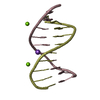
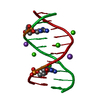
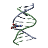
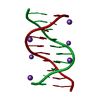
| Title | Crystal structure of d(CCGGTACCGG)2 as B-DNA duplex grown with 5 mM CoCl2 | ||||||||||||||||||
 Components Components | DNA (5'-D(* Keywords KeywordsDNA / Duplex | Function / homology | : / DNA |  Function and homology information Function and homology informationMethod |  X-RAY DIFFRACTION / SYNCHROTRON / MOLECULAR REPLACEMENT / Resolution: 2.8 Å X-RAY DIFFRACTION / SYNCHROTRON / MOLECULAR REPLACEMENT / Resolution: 2.8 Å  Authors AuthorsVenkadesh, S. / Mandal, P.K. / Gautham, N. |  CitationJournal: To be Published CitationJournal: To be PublishedTitle: Crystal structure of d(CCGGTACCGG)2 as B-DNA duplex Authors: Venkadesh, S. / Mandal, P.K. / Gautham, N. History |
|
- Structure visualization
| Structure viewer | Molecule: MolmilJmol/JSmol |
|---|
- Downloads & links
-Download
| PDBx/mmCIF format | 3r86.cif.gz | 22.9 KB | Display | PDBx/mmCIF format |
|---|---|---|---|---|
| PDB format | pdb3r86.ent.gz | 13.9 KB | Display | PDB format |
| PDBx/mmJSON format | 3r86.json.gz | Tree view | PDBx/mmJSON format | |
| Others |  Other downloads Other downloads |
-Validation report
| Arichive directory | https://data.pdbj.org/pub/pdb/validation_reports/r8/3r86ftp://data.pdbj.org/pub/pdb/validation_reports/r8/3r86 | HTTPS FTP |
|---|
-Related structure data
| Similar structure data |
|---|


-Links
 PDBj
PDBj


- Assembly
| Deposited unit | 
| ||||||||
|---|---|---|---|---|---|---|---|---|---|
| 1 |
| ||||||||
| Unit cell |
|
-Components
| #1: DNA chain | Mass: 3045.992 Da / Num. of mol.: 2 / Source method: obtained synthetically / Details: Chemically synthesized #2: Chemical |   Mass: 58.933 Da / Num. of mol.: 3 / Source method: obtained synthetically / Formula: Co Mass: 58.933 Da / Num. of mol.: 3 / Source method: obtained synthetically / Formula: Co#3: Water | ChemComp-HOH / |  Mass: 18.015 Da / Num. of mol.: 11 / Source method: isolated from a natural source / Formula: H2O Mass: 18.015 Da / Num. of mol.: 11 / Source method: isolated from a natural source / Formula: H2O |
|---|
-Experimental details
-Experiment
| Experiment | Method: X-RAY DIFFRACTION / Number of used crystals: 1 |
|---|
- Sample preparation
| Crystal | Density Matthews: 2.38 Å3/Da / Density % sol: 48.33 % |
|---|---|
| Crystal grow | Temperature: 293 K / Method: vapor diffusion, hanging drop / pH: 7 Details: 1mM DNA, 50mM Sodium Cacodylate buffer, 5mM CoCl2,1mM Spermine, 50% MPD, pH 7.0, VAPOR DIFFUSION, HANGING DROP, temperature 293K |
-Data collection
| Diffraction | Mean temperature: 100 K | |||||||||||||||
|---|---|---|---|---|---|---|---|---|---|---|---|---|---|---|---|---|
| Diffraction source | Source: SYNCHROTRON / Site: ESRF  / Beamline: BM14 / Wavelength: 1.604 Å / Beamline: BM14 / Wavelength: 1.604 Å | |||||||||||||||
| Detector | Type: MARMOSAIC 225 mm CCD / Detector: CCD / Date: Jul 7, 2010 | |||||||||||||||
| Radiation | Monochromator: Si(111) monochromator / Protocol: SINGLE WAVELENGTH / Monochromatic (M) / Laue (L): M / Scattering type: x-ray | |||||||||||||||
| Radiation wavelength | Wavelength: 1.604 Å / Relative weight: 1 | |||||||||||||||
| Reflection twin |
| |||||||||||||||
| Reflection | Resolution: 2.8→28.4 Å / Num. all: 1410 / Num. obs: 1382 / % possible obs: 98 % / Observed criterion σ(F): 1 / Observed criterion σ(I): 1 / Redundancy: 5.4 % / Biso Wilson estimate: 67.3 Å2 / Rmerge(I) obs: 0.051 / Rsym value: 0.047 / Net I/σ(I): 3.7 | |||||||||||||||
| Reflection shell | Resolution: 2.803→2.873 Å / Redundancy: 5.5 % / Rmerge(I) obs: 0.39 / Mean I/σ(I) obs: 1.4 / Num. unique all: 77 / Rsym value: 0.402 / % possible all: 99 |
- Processing
| Software |
| |||||||||||||||||||||||||
|---|---|---|---|---|---|---|---|---|---|---|---|---|---|---|---|---|---|---|---|---|---|---|---|---|---|---|
| Refinement | Method to determine structure: MOLECULAR REPLACEMENT Starting model: B-DNA fiber model Resolution: 2.8→28.37 Å / Cor.coef. Fo:Fc: 0.95 / Cor.coef. Fo:Fc free: 0.934 / SU B: 21.376 / SU ML: 0.384 / Isotropic thermal model: isotropic / Cross valid method: THROUGHOUT / ESU R Free: 0.105 / Stereochemistry target values: MAXIMUM LIKELIHOOD
| |||||||||||||||||||||||||
| Solvent computation | Ion probe radii: 0.8 Å / Shrinkage radii: 0.8 Å / VDW probe radii: 1.4 Å / Solvent model: BABINET MODEL WITH MASK | |||||||||||||||||||||||||
| Displacement parameters | Biso mean: 45.978 Å2
| |||||||||||||||||||||||||
| Refinement step | Cycle: LAST / Resolution: 2.8→28.37 Å
| |||||||||||||||||||||||||
| Refine LS restraints |
| |||||||||||||||||||||||||
| LS refinement shell | Resolution: 2.803→2.875 Å / Total num. of bins used: 20
|
