cellular response to nitroglycerin / response to hypobaric hypoxia / sequence-specific single stranded DNA binding / cellular response to diamide / cellular response to L-glutamine / negative regulation of double-strand break repair via nonhomologous end joining / positive regulation of stress granule assembly / positive regulation of apoptotic DNA fragmentation / translation elongation factor binding / negative regulation of inclusion body assembly ...cellular response to nitroglycerin / response to hypobaric hypoxia / sequence-specific single stranded DNA binding / cellular response to diamide / cellular response to L-glutamine / negative regulation of double-strand break repair via nonhomologous end joining / positive regulation of stress granule assembly / positive regulation of apoptotic DNA fragmentation / translation elongation factor binding / negative regulation of inclusion body assembly / positive regulation of inclusion body assembly / nuclear stress granule / cellular response to sodium arsenite / cellular response to potassium ion / positive regulation of macrophage differentiation / cellular response to angiotensin / protein folding chaperone complex / negative regulation of cardiac muscle cell apoptotic process / response to psychosocial stress / STAT family protein binding / RNA polymerase II intronic transcription regulatory region sequence-specific DNA binding / response to testosterone / mitotic spindle pole / general transcription initiation factor binding / HSF1-dependent transactivation / Regulation of HSF1-mediated heat shock response / HSF1 activation / Attenuation phase / cellular response to unfolded protein / mRNA transport / negative regulation of protein-containing complex assembly / heterochromatin / regulation of cellular response to heat / positive regulation of tyrosine phosphorylation of STAT protein / cellular response to copper ion / heat shock protein binding / cellular response to cadmium ion / positive regulation of mitotic cell cycle / response to nutrient / response to activity / cellular response to estradiol stimulus / promoter-specific chromatin binding / Hsp90 protein binding / euchromatin / chromatin DNA binding / cellular response to gamma radiation / mRNA processing / PML body / kinetochore / defense response / positive regulation of DNA-binding transcription factor activity / DNA-binding transcription repressor activity, RNA polymerase II-specific / Aggrephagy / cellular response to hydrogen peroxide / : / MAPK cascade / sequence-specific double-stranded DNA binding / cellular response to xenobiotic stimulus / positive regulation of cold-induced thermogenesis / cellular response to heat / protein-containing complex assembly / DNA-binding transcription activator activity, RNA polymerase II-specific / cellular response to lipopolysaccharide / sequence-specific DNA binding / transcription cis-regulatory region binding / DNA-binding transcription factor activity, RNA polymerase II-specific / ribonucleoprotein complex / protein heterodimerization activity / RNA polymerase II cis-regulatory region sequence-specific DNA binding / DNA-binding transcription factor activity / negative regulation of gene expression / DNA repair / centrosome / positive regulation of gene expression / chromatin / regulation of transcription by RNA polymerase II / protein kinase binding / perinuclear region of cytoplasm / negative regulation of transcription by RNA polymerase II / positive regulation of transcription by RNA polymerase II / DNA binding / nucleoplasm / identical protein binding / nucleus / cytoplasm / cytosol Similarity search - Function
In the structure databanks used in Yorodumi, some data are registered as the other names, "COVID-19 virus" and "2019-nCoV". Here are the details of the virus and the list of structure data.
Jan 31, 2019. EMDB accession codes are about to change! (news from PDBe EMDB page)
EMDB accession codes are about to change! (news from PDBe EMDB page)
The allocation of 4 digits for EMDB accession codes will soon come to an end. Whilst these codes will remain in use, new EMDB accession codes will include an additional digit and will expand incrementally as the available range of codes is exhausted. The current 4-digit format prefixed with “EMD-” (i.e. EMD-XXXX) will advance to a 5-digit format (i.e. EMD-XXXXX), and so on. It is currently estimated that the 4-digit codes will be depleted around Spring 2019, at which point the 5-digit format will come into force.
The EM Navigator/Yorodumi systems omit the EMD- prefix.
Related info.:Q: What is EMD? / ID/Accession-code notation in Yorodumi/EM Navigator
Yorodumi is a browser for structure data from EMDB, PDB, SASBDB, etc.
This page is also the successor to EM Navigator detail page, and also detail information page/front-end page for Omokage search.
The word "yorodu" (or yorozu) is an old Japanese word meaning "ten thousand". "mi" (miru) is to see.
Related info.:EMDB / PDB / SASBDB / Comparison of 3 databanks / Yorodumi Search / Aug 31, 2016. New EM Navigator & Yorodumi / Yorodumi Papers / Jmol/JSmol / Function and homology information / Changes in new EM Navigator and Yorodumi
 Movie
Movie Controller


 Open data
 Basic information
 Components
 Keywords
 Function and homology information
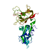
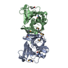
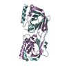
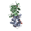
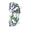
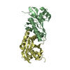
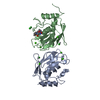
 Homo sapiens (human)
 X-RAY DIFFRACTION /
  Authors
 Citation
 Structure visualization
 Downloads & links
 Other downloads
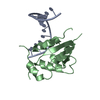


 PDBj
PDBj


 Assembly


 Mass: 24.305 Da / Num. of mol.: 2 / Source method: obtained synthetically / Formula: Mg
 Mass: 18.015 Da / Num. of mol.: 108 / Source method: isolated from a natural source / Formula: H2O
 Sample preparation
 / Beamline: ID23-2 / Wavelength: 0.8726 Å
 Processing