 Movie
Movie Controller


+ Open data

- Basic information
| Entry | Database: PDB / ID: 5d5x | ||||||
|---|---|---|---|---|---|---|---|
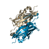
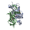
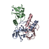
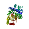
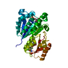
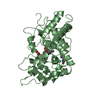
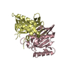
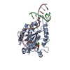
| Title | Crystal structure of Chaetomium thermophilum Skn7 with SSRE DNA | ||||||
 Components Components |
| ||||||
 Keywords Keywords | TRANSCRIPTION / protein-DNA complex / double helix / helix-turn-helix | ||||||
| Function / homology |  Function and homology information Function and homology informationphosphorelay signal transduction system / sequence-specific DNA binding / DNA-binding transcription factor activity / identical protein binding / nucleus Similarity search - Function | ||||||
| Biological species |  Chaetomium thermophilum (fungus) Chaetomium thermophilum (fungus) | ||||||
| Method |  X-RAY DIFFRACTION / SYNCHROTRON / MOLECULAR REPLACEMENT / molecular replacement / Resolution: 2.4 Å X-RAY DIFFRACTION / SYNCHROTRON / MOLECULAR REPLACEMENT / molecular replacement / Resolution: 2.4 Å | ||||||
 Authors Authors | Neudegger, T. / Verghese, J. / Hayer-Hartl, M. / Hartl, F.U. / Bracher, A. | ||||||
 Citation Citation | Journal: Nat.Struct.Mol.Biol. / Year: 2016 Title: Structure of human heat-shock transcription factor 1 in complex with DNA. Authors: Neudegger, T. / Verghese, J. / Hayer-Hartl, M. / Hartl, F.U. / Bracher, A. | ||||||
| History |
|
- Structure visualization
| Structure viewer | Molecule: MolmilJmol/JSmol |
|---|
- Downloads & links
-Download
| PDBx/mmCIF format | 5d5x.cif.gz | 72 KB | Display | PDBx/mmCIF format |
|---|---|---|---|---|
| PDB format | pdb5d5x.ent.gz | 50.1 KB | Display | PDB format |
| PDBx/mmJSON format | 5d5x.json.gz | Tree view | PDBx/mmJSON format | |
| Others |  Other downloads Other downloads |
-Validation report
| Arichive directory | https://data.pdbj.org/pub/pdb/validation_reports/d5/5d5xftp://data.pdbj.org/pub/pdb/validation_reports/d5/5d5x | HTTPS FTP |
|---|
-Related structure data
| Related structure data | 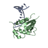 5d5uC 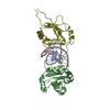 5d5vC  5d5wSC  5d5yC  5d5zC  5d60C S: Starting model for refinement C: citing same article ( |
|---|---|
| Similar structure data |


-Links
 PDBj
PDBj


- Assembly
| Deposited unit | 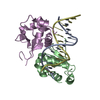
| ||||||||
|---|---|---|---|---|---|---|---|---|---|
| 1 |
| ||||||||
| Unit cell |
| ||||||||
| Components on special symmetry positions |
|
-Components
| #1: DNA chain | Mass: 3945.589 Da / Num. of mol.: 1 / Fragment: DNA binding domain / Source method: obtained synthetically / Source: (synth.) | ||
|---|---|---|---|
| #2: DNA chain | Mass: 3998.594 Da / Num. of mol.: 1 / Source method: obtained synthetically / Source: (synth.) | ||
| #3: Protein | Mass: 12618.037 Da / Num. of mol.: 2 Source method: isolated from a genetically manipulated source Source: (gene. exp.) Chaetomium thermophilum (fungus) / Gene: CTHT_0048700 / Plasmid: pHUE / Production host:  #4: Water | ChemComp-HOH / |  Mass: 18.015 Da / Num. of mol.: 39 / Source method: isolated from a natural source / Formula: H2O Mass: 18.015 Da / Num. of mol.: 39 / Source method: isolated from a natural source / Formula: H2O |
-Experimental details
-Experiment
| Experiment | Method: X-RAY DIFFRACTION / Number of used crystals: 1 |
|---|
- Sample preparation
| Crystal | Density Matthews: 2.13 Å3/Da / Density % sol: 42.38 % |
|---|---|
| Crystal grow | Temperature: 293 K / Method: vapor diffusion, hanging drop / pH: 7.5 / Details: 25 % PEG-2000 MME, 0.1 M HEPES-NaOH pH 7.5 |
-Data collection
| Diffraction | Mean temperature: 100 K | |||||||||||||||||||||||||||
|---|---|---|---|---|---|---|---|---|---|---|---|---|---|---|---|---|---|---|---|---|---|---|---|---|---|---|---|---|
| Diffraction source | Source: SYNCHROTRON / Site: ESRF  / Beamline: MASSIF-1 / Wavelength: 0.965 Å / Beamline: MASSIF-1 / Wavelength: 0.965 Å | |||||||||||||||||||||||||||
| Detector | Type: DECTRIS PILATUS 2M / Detector: PIXEL / Date: Nov 5, 2014 | |||||||||||||||||||||||||||
| Radiation | Protocol: SINGLE WAVELENGTH / Monochromatic (M) / Laue (L): M / Scattering type: x-ray | |||||||||||||||||||||||||||
| Radiation wavelength | Wavelength: 0.965 Å / Relative weight: 1 | |||||||||||||||||||||||||||
| Reflection | Resolution: 2.39→44.34 Å / Num. obs: 11227 / % possible obs: 99.3 % / Redundancy: 5.1 % / Biso Wilson estimate: 45.5 Å2 / CC1/2: 0.997 / Rmerge(I) obs: 0.092 / Rpim(I) all: 0.045 / Net I/σ(I): 10.5 / Num. measured all: 56918 | |||||||||||||||||||||||||||
| Reflection shell | Diffraction-ID: 1 / Rejects: _
|
-Phasing
| Phasing | Method: molecular replacement |
|---|
- Processing
| Software |
| ||||||||||||||||||||||||||||||||||||||||||||||||||||||||||||
|---|---|---|---|---|---|---|---|---|---|---|---|---|---|---|---|---|---|---|---|---|---|---|---|---|---|---|---|---|---|---|---|---|---|---|---|---|---|---|---|---|---|---|---|---|---|---|---|---|---|---|---|---|---|---|---|---|---|---|---|---|---|
| Refinement | Method to determine structure: MOLECULAR REPLACEMENT Starting model: 5D5W Resolution: 2.4→30 Å / Cor.coef. Fo:Fc: 0.939 / Cor.coef. Fo:Fc free: 0.9 / WRfactor Rfree: 0.2814 / WRfactor Rwork: 0.2249 / FOM work R set: 0.7616 / SU B: 11.464 / SU ML: 0.269 / SU R Cruickshank DPI: 0.6397 / SU Rfree: 0.3232 / Cross valid method: THROUGHOUT / σ(F): 0 / ESU R: 0.64 / ESU R Free: 0.323 / Stereochemistry target values: MAXIMUM LIKELIHOOD Details: HYDROGENS HAVE BEEN ADDED IN THE RIDING POSITIONS U VALUES : REFINED INDIVIDUALLY
| ||||||||||||||||||||||||||||||||||||||||||||||||||||||||||||
| Solvent computation | Ion probe radii: 0.8 Å / Shrinkage radii: 0.8 Å / VDW probe radii: 1.2 Å / Solvent model: MASK | ||||||||||||||||||||||||||||||||||||||||||||||||||||||||||||
| Displacement parameters | Biso max: 112.52 Å2 / Biso mean: 50.29 Å2 / Biso min: 23.96 Å2
| ||||||||||||||||||||||||||||||||||||||||||||||||||||||||||||
| Refinement step | Cycle: final / Resolution: 2.4→30 Å
| ||||||||||||||||||||||||||||||||||||||||||||||||||||||||||||
| Refine LS restraints |
| ||||||||||||||||||||||||||||||||||||||||||||||||||||||||||||
| LS refinement shell | Resolution: 2.4→2.462 Å / Total num. of bins used: 20
|
