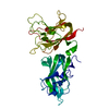
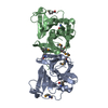
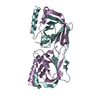
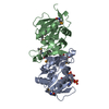
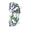
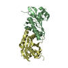
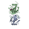
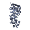
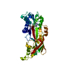
Entry Database : PDB / ID : 5d5vTitle Crystal structure of human Hsf1 with Satellite III repeat DNA (DNA) x 2 Heat shock factor protein 1 Keywords / / / / Function / homology Function Domain/homology Component
/ / / / / / / / / / / / / / / / / / / / / / / / / / / / / / / / / / / / / / / / / / / / / / / / / / / / / / / / / / / / / / / / / / / / / / / / / / / / / / / / / / / / / / / / / / / / / / / / / / / / / / / / / / / / / / / / / / / / / / / / / / Biological species Homo sapiens (human)Method / / / / Resolution : 2.55 Å Authors Neudegger, T. / Verghese, J. / Hayer-Hartl, M. / Hartl, F.U. / Bracher, A. Journal : Nat.Struct.Mol.Biol. / Year : 2016Title : Structure of human heat-shock transcription factor 1 in complex with DNA.Authors : Neudegger, T. / Verghese, J. / Hayer-Hartl, M. / Hartl, F.U. / Bracher, A. History Deposition Aug 11, 2015 Deposition site / Processing site Revision 1.0 Dec 30, 2015 Provider / Type Revision 1.1 Jan 13, 2016 Group Revision 1.2 Jan 20, 2016 Group Revision 1.3 Feb 10, 2016 Group Revision 1.4 Jan 10, 2024 Group / Database references / Refinement descriptionCategory chem_comp_atom / chem_comp_bond ... chem_comp_atom / chem_comp_bond / database_2 / diffrn_radiation_wavelength / pdbx_initial_refinement_model / struct_ncs_dom_lim Item _database_2.pdbx_DOI / _database_2.pdbx_database_accession ... _database_2.pdbx_DOI / _database_2.pdbx_database_accession / _struct_ncs_dom_lim.beg_auth_comp_id / _struct_ncs_dom_lim.beg_label_asym_id / _struct_ncs_dom_lim.beg_label_comp_id / _struct_ncs_dom_lim.beg_label_seq_id / _struct_ncs_dom_lim.end_auth_comp_id / _struct_ncs_dom_lim.end_label_asym_id / _struct_ncs_dom_lim.end_label_comp_id / _struct_ncs_dom_lim.end_label_seq_id
Show all Show less
 Movie
Movie Controller


 Open data
 Basic information
 Components
 Keywords
 Function and homology information
 Homo sapiens (human)
 X-RAY DIFFRACTION /
  Authors
 Citation
 Structure visualization
 Downloads & links
 Other downloads
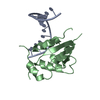


 PDBj
PDBj


 Assembly
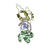


 Mass: 24.305 Da / Num. of mol.: 2 / Source method: obtained synthetically / Formula: Mg
 Mass: 18.015 Da / Num. of mol.: 108 / Source method: isolated from a natural source / Formula: H2O
 Sample preparation
 / Beamline: ID23-2 / Wavelength: 0.8726 Å
 Processing