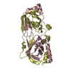
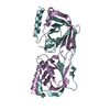
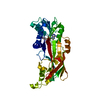
BIOMOLECULE: 1, 2 SEE REMARK 350 FOR THE AUTHOR PROVIDED AND PROGRAM GENERATED ASSEMBLY ... BIOMOLECULE: 1, 2 SEE REMARK 350 FOR THE AUTHOR PROVIDED AND PROGRAM GENERATED ASSEMBLY INFORMATION FOR THE STRUCTURE IN THIS ENTRY. THE REMARK MAY ALSO PROVIDE INFORMATION ON BURIED SURFACE AREA. SIZE EXCLUSION CHROMATOGRAPHY SUPPORTS THE ASSIGNMENT OF A DIMER AS THE SIGNIFICANT OLIGOMERIZATION STATE.
Remark 999
SEQUENCE THE CONSTRUCT WAS EXPRESSED WITH A PURIFICATION TAG MGSDKIHHHHHHENLYFQG WHICH WAS NOT ... SEQUENCE THE CONSTRUCT WAS EXPRESSED WITH A PURIFICATION TAG MGSDKIHHHHHHENLYFQG WHICH WAS NOT CLEAVED AFTER PURIFICATION.
Monochromator: Single crystal Si(111) bent (horizontal focusing) Protocol: MAD / Monochromatic (M) / Laue (L): M / Scattering type: x-ray
Radiation wavelength
ID
Wavelength (Å)
Relative weight
1
0.91837
1
2
0.97886
1
3
0.97917
1
Reflection
Resolution: 1.6→29.54 Å / Num. obs: 75349 / % possible obs: 99.2 % / Observed criterion σ(I): -3 / Biso Wilson estimate: 20.872 Å2 / Rmerge(I) obs: 0.053 / Net I/σ(I): 10.8
Reflection shell
Resolution (Å)
Rmerge(I) obs
Mean I/σ(I) obs
Num. measured obs
Num. unique obs
% possible all
1.6-1.66
0.368
2.2
23229
14986
98.5
1.66-1.72
0.303
2.7
20768
13243
99.8
1.72-1.8
0.246
3.3
23340
14854
99.7
1.8-1.9
0.177
4.5
24353
15308
99.7
1.9-2.02
0.128
6.2
23292
14535
99.5
2.02-2.17
0.089
8.7
22446
13902
99.5
2.17-2.39
0.064
11.7
23905
14603
99.6
2.39-2.73
0.049
14.9
23733
14332
99.5
2.73-3.44
0.031
21.5
24419
14582
99.3
3.44-29.54
0.02
32.7
24734
14311
97.4
-
Phasing
Phasing
Method: MAD
-
Processing
Software
Name
Version
Classification
NB
REFMAC
5.3.0040
refinement
PHENIX
refinement
SOLVE
phasing
MolProbity
3beta29
modelbuilding
XSCALE
datascaling
PDB_EXTRACT
3
dataextraction
MAR345
CCD
datacollection
XDS
datareduction
Refinement
Method to determine structure: MAD / Resolution: 1.6→29.54 Å / Cor.coef. Fo:Fc: 0.967 / Cor.coef. Fo:Fc free: 0.95 / SU B: 1.444 / SU ML: 0.052 / Cross valid method: THROUGHOUT / σ(F): 0 / ESU R: 0.079 / ESU R Free: 0.082 Stereochemistry target values: MAXIMUM LIKELIHOOD WITH PHASES Details: 1. HYDROGENS HAVE BEEN ADDED IN THE RIDING POSITIONS. 2. A MET-INHIBITION PROTOCOL WAS USED FOR SELENOMETHIONINE INCORPORATION DURING PROTEIN EXPRESSION. THE OCCUPANCY OF THE SE ATOMS IN THE ...Details: 1. HYDROGENS HAVE BEEN ADDED IN THE RIDING POSITIONS. 2. A MET-INHIBITION PROTOCOL WAS USED FOR SELENOMETHIONINE INCORPORATION DURING PROTEIN EXPRESSION. THE OCCUPANCY OF THE SE ATOMS IN THE MSE RESIDUES WAS REDUCED TO 0.75 TO ACCOUNT FOR THE REDUCED SCATTERING POWER DUE TO PARTIAL S-MET INCORPORATION. 3. ONE PEG MOLECULE HAS BEEN MODELED IN THE SOLVENT STRUCTURE.
Rfactor
Num. reflection
% reflection
Selection details
Rfree
0.186
3791
5 %
RANDOM
Rwork
0.151
-
-
-
obs
0.153
75306
99.59 %
-
Solvent computation
Ion probe radii: 0.8 Å / Shrinkage radii: 0.8 Å / VDW probe radii: 1.2 Å / Solvent model: MASK
Displacement parameters
Biso mean: 11.56 Å2
Baniso -1
Baniso -2
Baniso -3
1-
0.29 Å2
0 Å2
0 Å2
2-
-
-0.14 Å2
0 Å2
3-
-
-
-0.15 Å2
Refinement step
Cycle: LAST / Resolution: 1.6→29.54 Å
Protein
Nucleic acid
Ligand
Solvent
Total
Num. atoms
4543
0
48
626
5217
Refine LS restraints
Refine-ID
Type
Dev ideal
Dev ideal target
Number
X-RAY DIFFRACTION
r_bond_refined_d
0.017
0.022
4704
X-RAY DIFFRACTION
r_bond_other_d
0.004
0.02
3039
X-RAY DIFFRACTION
r_angle_refined_deg
1.751
1.966
6420
X-RAY DIFFRACTION
r_angle_other_deg
1.42
3
7535
X-RAY DIFFRACTION
r_dihedral_angle_1_deg
4.599
5
630
X-RAY DIFFRACTION
r_dihedral_angle_2_deg
40.163
26.2
200
X-RAY DIFFRACTION
r_dihedral_angle_3_deg
10.461
15
796
X-RAY DIFFRACTION
r_dihedral_angle_4_deg
7.544
15
10
X-RAY DIFFRACTION
r_chiral_restr
0.086
0.2
736
X-RAY DIFFRACTION
r_gen_planes_refined
0.007
0.02
5315
X-RAY DIFFRACTION
r_gen_planes_other
0.003
0.02
859
X-RAY DIFFRACTION
r_nbd_refined
0.2
0.3
870
X-RAY DIFFRACTION
r_nbd_other
0.157
0.3
3146
X-RAY DIFFRACTION
r_nbtor_refined
0.175
0.5
2293
X-RAY DIFFRACTION
r_nbtor_other
0.085
0.5
2301
X-RAY DIFFRACTION
r_xyhbond_nbd_refined
0.157
0.5
836
X-RAY DIFFRACTION
r_xyhbond_nbd_other
0.134
0.5
1
X-RAY DIFFRACTION
r_symmetry_vdw_refined
0.198
0.3
26
X-RAY DIFFRACTION
r_symmetry_vdw_other
0.239
0.3
65
X-RAY DIFFRACTION
r_symmetry_hbond_refined
0.175
0.5
78
X-RAY DIFFRACTION
r_mcbond_it
2.227
3
3239
X-RAY DIFFRACTION
r_mcbond_other
0.532
3
1224
X-RAY DIFFRACTION
r_mcangle_it
2.789
5
4799
X-RAY DIFFRACTION
r_scbond_it
4.512
8
1884
X-RAY DIFFRACTION
r_scangle_it
6.387
11
1600
LS refinement shell
Resolution: 1.6→1.642 Å / Total num. of bins used: 20
Rfactor
Num. reflection
% reflection
Rfree
0.254
277
-
Rwork
0.187
5170
-
all
-
5447
-
obs
-
-
99.22 %
+
About Yorodumi
-
News
-
Feb 9, 2022. New format data for meta-information of EMDB entries
New format data for meta-information of EMDB entries
Version 3 of the EMDB header file is now the official format.
The previous official version 1.9 will be removed from the archive.
In the structure databanks used in Yorodumi, some data are registered as the other names, "COVID-19 virus" and "2019-nCoV". Here are the details of the virus and the list of structure data.
Jan 31, 2019. EMDB accession codes are about to change! (news from PDBe EMDB page)
EMDB accession codes are about to change! (news from PDBe EMDB page)
The allocation of 4 digits for EMDB accession codes will soon come to an end. Whilst these codes will remain in use, new EMDB accession codes will include an additional digit and will expand incrementally as the available range of codes is exhausted. The current 4-digit format prefixed with “EMD-” (i.e. EMD-XXXX) will advance to a 5-digit format (i.e. EMD-XXXXX), and so on. It is currently estimated that the 4-digit codes will be depleted around Spring 2019, at which point the 5-digit format will come into force.
The EM Navigator/Yorodumi systems omit the EMD- prefix.
Related info.:Q: What is EMD? / ID/Accession-code notation in Yorodumi/EM Navigator
Yorodumi is a browser for structure data from EMDB, PDB, SASBDB, etc.
This page is also the successor to EM Navigator detail page, and also detail information page/front-end page for Omokage search.
The word "yorodu" (or yorozu) is an old Japanese word meaning "ten thousand". "mi" (miru) is to see.
Related info.:EMDB / PDB / SASBDB / Comparison of 3 databanks / Yorodumi Search / Aug 31, 2016. New EM Navigator & Yorodumi / Yorodumi Papers / Jmol/JSmol / Function and homology information / Changes in new EM Navigator and Yorodumi
 Movie
Movie Controller

 Yorodumi
 Open data
 Basic information
 Components
 Keywords
 Function and homology information
 Lactobacillus plantarum WCFS1 (bacteria)
 X-RAY DIFFRACTION /
  Authors
 Citation
 Structure visualization
 Downloads & links
 Other downloads
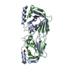


 PDBj
PDBj Assembly

 Mass: 743.405 Da / Num. of mol.: 1 / Source method: obtained synthetically / Formula: C21H28N7O17P3
 Mass: 106.120 Da / Num. of mol.: 1 / Source method: obtained synthetically / Formula: C4H10O3
 Mass: 18.015 Da / Num. of mol.: 619 / Source method: isolated from a natural source / Formula: H2O
 Sample preparation
 / Beamline: BL11-1 / Wavelength: 0.91837, 0.97886, 0.97917
 Processing