 Movie
Movie Controller

[English] 日本語
 Yorodumi
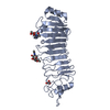
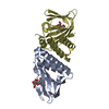
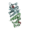
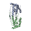
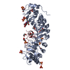
- PDB-6i6j: Crystal structure of the KDEL receptor bound to synthetic nanobody. -
+ Open data

- Basic information
| Entry | Database: PDB / ID: 6i6j | ||||||
|---|---|---|---|---|---|---|---|
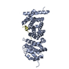
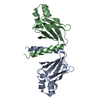
| Title | Crystal structure of the KDEL receptor bound to synthetic nanobody. | ||||||
 Components Components |
| ||||||
 Keywords Keywords | MEMBRANE PROTEIN / Intracellular protein receptor / KDEL / ERD2 | ||||||
| Function / homology |  Function and homology information Function and homology informationKDEL sequence binding / COPI-coated vesicle membrane / ER lumen protein retrieval receptor activity / COPI-dependent Golgi-to-ER retrograde traffic / COPI-mediated anterograde transport / protein retention in ER lumen / maintenance of protein localization in endoplasmic reticulum / cis-Golgi network / retrograde vesicle-mediated transport, Golgi to endoplasmic reticulum / protein transport ...KDEL sequence binding / COPI-coated vesicle membrane / ER lumen protein retrieval receptor activity / COPI-dependent Golgi-to-ER retrograde traffic / COPI-mediated anterograde transport / protein retention in ER lumen / maintenance of protein localization in endoplasmic reticulum / cis-Golgi network / retrograde vesicle-mediated transport, Golgi to endoplasmic reticulum / protein transport / Golgi membrane / endoplasmic reticulum membrane / endoplasmic reticulum / membrane Similarity search - Function | ||||||
| Biological species |  synthetic construct (others) | ||||||
| Method |  X-RAY DIFFRACTION / SYNCHROTRON / MOLECULAR REPLACEMENT / Resolution: 2.23 Å X-RAY DIFFRACTION / SYNCHROTRON / MOLECULAR REPLACEMENT / Resolution: 2.23 Å | ||||||
 Authors Authors | Braeuer, P. / Newstead, S. | ||||||
| Funding support |  United Kingdom, 1items United Kingdom, 1items
| ||||||
 Citation Citation | Journal: Science / Year: 2019 Title: Structural basis for pH-dependent retrieval of ER proteins from the Golgi by the KDEL receptor. Authors: Brauer, P. / Parker, J.L. / Gerondopoulos, A. / Zimmermann, I. / Seeger, M.A. / Barr, F.A. / Newstead, S. | ||||||
| History |
|
- Structure visualization
| Structure viewer | Molecule: MolmilJmol/JSmol |
|---|
- Downloads & links
-Download
| PDBx/mmCIF format | 6i6j.cif.gz | 149.6 KB | Display | PDBx/mmCIF format |
|---|---|---|---|---|
| PDB format | pdb6i6j.ent.gz | 116 KB | Display | PDB format |
| PDBx/mmJSON format | 6i6j.json.gz | Tree view | PDBx/mmJSON format | |
| Others |  Other downloads Other downloads |
-Validation report
| Arichive directory | https://data.pdbj.org/pub/pdb/validation_reports/i6/6i6jftp://data.pdbj.org/pub/pdb/validation_reports/i6/6i6j | HTTPS FTP |
|---|
-Related structure data
| Related structure data |  6i6bC 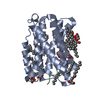 6i6hC 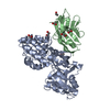 5m13S C: citing same article ( S: Starting model for refinement |
|---|---|
| Similar structure data |


-Links
 PDBj
PDBj


- Assembly
| Deposited unit | 
| ||||||||
|---|---|---|---|---|---|---|---|---|---|
| 1 |
| ||||||||
| Unit cell |
|
-Components
| #1: Protein | Mass: 23679.887 Da / Num. of mol.: 1 Source method: isolated from a genetically manipulated source Source: (gene. exp.)  | ||||
|---|---|---|---|---|---|
| #2: Antibody | Mass: 13611.107 Da / Num. of mol.: 1 Source method: isolated from a genetically manipulated source Source: (gene. exp.) synthetic construct (others) / Production host:  | ||||
| #3: Chemical |   Mass: 356.540 Da / Num. of mol.: 2 / Source method: obtained synthetically / Formula: C21H40O4 Mass: 356.540 Da / Num. of mol.: 2 / Source method: obtained synthetically / Formula: C21H40O4#4: Water | ChemComp-HOH / |  Mass: 18.015 Da / Num. of mol.: 79 / Source method: isolated from a natural source / Formula: H2O Mass: 18.015 Da / Num. of mol.: 79 / Source method: isolated from a natural source / Formula: H2OHas protein modification | Y | |
-Experimental details
-Experiment
| Experiment | Method: X-RAY DIFFRACTION / Number of used crystals: 1 |
|---|
- Sample preparation
| Crystal | Density Matthews: 2.38 Å3/Da / Density % sol: 48.36 % |
|---|---|
| Crystal grow | Temperature: 293 K / Method: lipidic cubic phase / pH: 9 / Details: 30% (v/v) PEG 400, 100 mM Tris pH 9.0 |
-Data collection
| Diffraction | Mean temperature: 100 K / Serial crystal experiment: N | ||||||||||||||||||||||||
|---|---|---|---|---|---|---|---|---|---|---|---|---|---|---|---|---|---|---|---|---|---|---|---|---|---|
| Diffraction source | Source: SYNCHROTRON / Site: Diamond / Beamline: I24 / Wavelength: 0.968 Å | ||||||||||||||||||||||||
| Detector | Type: DECTRIS PILATUS3 6M / Detector: PIXEL / Date: Jun 27, 2018 | ||||||||||||||||||||||||
| Radiation | Protocol: SINGLE WAVELENGTH / Monochromatic (M) / Laue (L): M / Scattering type: x-ray | ||||||||||||||||||||||||
| Radiation wavelength | Wavelength: 0.968 Å / Relative weight: 1 | ||||||||||||||||||||||||
| Reflection | Resolution: 2.23→52.97 Å / Num. obs: 18081 / % possible obs: 100 % / Redundancy: 6.2 % / CC1/2: 0.869 / Rmerge(I) obs: 0.122 / Rpim(I) all: 0.055 / Rrim(I) all: 0.134 / Net I/σ(I): 7 | ||||||||||||||||||||||||
| Reflection shell | Diffraction-ID: 1
|
- Processing
| Software |
| |||||||||||||||||||||||||||||||||||||||||||||||||||||||||||||||||||||||||||
|---|---|---|---|---|---|---|---|---|---|---|---|---|---|---|---|---|---|---|---|---|---|---|---|---|---|---|---|---|---|---|---|---|---|---|---|---|---|---|---|---|---|---|---|---|---|---|---|---|---|---|---|---|---|---|---|---|---|---|---|---|---|---|---|---|---|---|---|---|---|---|---|---|---|---|---|---|
| Refinement | Method to determine structure: MOLECULAR REPLACEMENT Starting model: 5M13 Resolution: 2.23→47.132 Å / SU ML: 0.22 / Cross valid method: THROUGHOUT / σ(F): 1.35 / Phase error: 24.57
| |||||||||||||||||||||||||||||||||||||||||||||||||||||||||||||||||||||||||||
| Solvent computation | Shrinkage radii: 0.9 Å / VDW probe radii: 1.11 Å | |||||||||||||||||||||||||||||||||||||||||||||||||||||||||||||||||||||||||||
| Displacement parameters | Biso max: 123.08 Å2 / Biso mean: 49.3756 Å2 / Biso min: 22.62 Å2 | |||||||||||||||||||||||||||||||||||||||||||||||||||||||||||||||||||||||||||
| Refinement step | Cycle: final / Resolution: 2.23→47.132 Å
| |||||||||||||||||||||||||||||||||||||||||||||||||||||||||||||||||||||||||||
| LS refinement shell | Refine-ID: X-RAY DIFFRACTION / Rfactor Rfree error: 0 / Total num. of bins used: 6
| |||||||||||||||||||||||||||||||||||||||||||||||||||||||||||||||||||||||||||
| Refinement TLS params. | Method: refined / Refine-ID: X-RAY DIFFRACTION
| |||||||||||||||||||||||||||||||||||||||||||||||||||||||||||||||||||||||||||
| Refinement TLS group |
|
