 Movie
Movie Controller


+ Open data

- Basic information
| Entry | Database: PDB / ID: 5d5g | ||||||
|---|---|---|---|---|---|---|---|
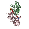
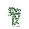
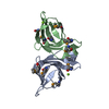
| Title | Structure of colocasia esculenta agglutinin | ||||||
 Components Components | (Tuber agglutinin) x 2 | ||||||
 Keywords Keywords | SUGAR BINDING PROTEIN / CARBOHYDRATE BINDING / LECTIN / AGGLUTININ / BETA PRISM II | ||||||
| Function / homology |  Function and homology information Function and homology informationresponse to other organism / D-mannose binding / extracellular region / metal ion binding Similarity search - Function | ||||||
| Biological species |  Colocasia esculenta (taro) Colocasia esculenta (taro) | ||||||
| Method |  X-RAY DIFFRACTION / MOLECULAR REPLACEMENT / Resolution: 1.74 Å X-RAY DIFFRACTION / MOLECULAR REPLACEMENT / Resolution: 1.74 Å | ||||||
 Authors Authors | Biswas, H. / Chattopadhyaya, R. | ||||||
| Funding support |  India, 1items India, 1items
| ||||||
 Citation Citation | Journal: J GLYCOBIO / Year: 2017 Title: Crystal structure Colocasia esculenta tuber agglutinin at 1.74A resolution and its quaternary interactions Authors: Chattopadhyaya, R. / Biswas, H. / Sarkar, A. #1: Journal: J. Biomol. Struct. Dyn. / Year: 2017 Title: Thermal and chemical denaturation of Colocasia esculenta tuber agglutinin from alpha 2 beta 2 to unfolded state Authors: Biswas, H. / Chattopadhyaya, R. | ||||||
| History |
|
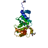
- Structure visualization
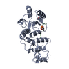
| Structure viewer | Molecule: MolmilJmol/JSmol |
|---|
- Downloads & links
-Download
| PDBx/mmCIF format | 5d5g.cif.gz | 103.3 KB | Display | PDBx/mmCIF format |
|---|---|---|---|---|
| PDB format | pdb5d5g.ent.gz | 78 KB | Display | PDB format |
| PDBx/mmJSON format | 5d5g.json.gz | Tree view | PDBx/mmJSON format | |
| Others |  Other downloads Other downloads |
-Validation report
| Arichive directory | https://data.pdbj.org/pub/pdb/validation_reports/d5/5d5gftp://data.pdbj.org/pub/pdb/validation_reports/d5/5d5g | HTTPS FTP |
|---|
-Related structure data
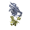
| Related structure data | 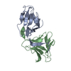 3r0eS S: Starting model for refinement |
|---|---|
| Similar structure data |


-Links
 PDBj
PDBj
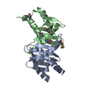
- Assembly
| Deposited unit | 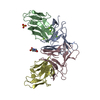
| ||||||||
|---|---|---|---|---|---|---|---|---|---|
| 1 | 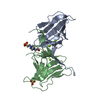
| ||||||||
| 2 | 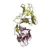
| ||||||||
| Unit cell |
|
-Components
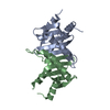
-Protein , 2 types, 4 molecules ACBD
| #1: Protein | Mass: 12012.415 Da / Num. of mol.: 2 / Fragment: UNP residues 24-132 / Source method: isolated from a natural source / Source: (natural) Colocasia esculenta (taro) / Plasmid details: local market, food item / Tissue: tuber / References: UniProt: R9RL27#2: Protein | Mass: 12418.863 Da / Num. of mol.: 2 / Fragment: UNP residues 140-250 / Source method: isolated from a natural source / Source: (natural) Colocasia esculenta (taro) / Plasmid details: local market, food item / Tissue: tuber / References: UniProt: R9RL27 |
|---|
-Non-polymers , 4 types, 141 molecules 


| #3: Chemical | ChemComp-MG /  Mass: 24.305 Da / Num. of mol.: 5 / Source method: obtained synthetically / Formula: Mg Mass: 24.305 Da / Num. of mol.: 5 / Source method: obtained synthetically / Formula: Mg#4: Chemical | ChemComp-EPE / |  Mass: 238.305 Da / Num. of mol.: 1 / Source method: obtained synthetically / Formula: C8H18N2O4S / Comment: pH buffer*YM Mass: 238.305 Da / Num. of mol.: 1 / Source method: obtained synthetically / Formula: C8H18N2O4S / Comment: pH buffer*YM#5: Chemical | ChemComp-SO4 / |  Mass: 96.063 Da / Num. of mol.: 1 / Source method: obtained synthetically / Formula: SO4 Mass: 96.063 Da / Num. of mol.: 1 / Source method: obtained synthetically / Formula: SO4#6: Water | ChemComp-HOH / | Mass: 18.015 Da / Num. of mol.: 134 / Source method: isolated from a natural source / Formula: H2O |
|---|
-Details
| Has protein modification | Y |
|---|
-Experimental details
-Experiment
| Experiment | Method: X-RAY DIFFRACTION / Number of used crystals: 1 |
|---|
- Sample preparation
| Crystal | Density Matthews: 2.42 Å3/Da / Density % sol: 49.27 % / Description: rectangular plates |
|---|---|
| Crystal grow | Temperature: 298 K / Method: vapor diffusion, hanging drop / pH: 6 Details: reservoir included 1.8 M ammonium sulphate and 0.1 M Hepes, pH 6.0; stock protein solution contained 8.1 mg/ml of lectin in 20 mM Tris pH 8.5; protein & reservoir soln mixed in 3:1 ratio at ...Details: reservoir included 1.8 M ammonium sulphate and 0.1 M Hepes, pH 6.0; stock protein solution contained 8.1 mg/ml of lectin in 20 mM Tris pH 8.5; protein & reservoir soln mixed in 3:1 ratio at start and placed on siliconized cover slip Temp details: fluctuated within 5 K |
-Data collection
| Diffraction | Mean temperature: 113 K / Ambient temp details: over 2 days, 8 min per frame |
|---|---|
| Diffraction source | Source: ROTATING ANODE / Type: RIGAKU RUH3R / Wavelength: 1.5419 Å |
| Detector | Type: RIGAKU RAXIS IV++ / Detector: IMAGE PLATE / Date: Dec 5, 2014 |
| Radiation | Monochromator: Ni filter / Protocol: SINGLE WAVELENGTH / Monochromatic (M) / Laue (L): M / Scattering type: x-ray |
| Radiation wavelength | Wavelength: 1.5419 Å / Relative weight: 1 |
| Reflection | Resolution: 1.536→21 Å / Num. obs: 59836 / % possible obs: 83.4 % / Redundancy: 5.61 % / Rmerge(I) obs: 0.38 / Rsym value: 0.38 / Net I/σ(I): 2.7 |
| Reflection shell | Resolution: 1.536→1.58 Å / Redundancy: 1.92 % / Rmerge(I) obs: 0.61 / Mean I/σ(I) obs: 0.9 / % possible all: 20 |
- Processing
| Software |
| |||||||||||||||||||||||||||||||||||||||||||||||||
|---|---|---|---|---|---|---|---|---|---|---|---|---|---|---|---|---|---|---|---|---|---|---|---|---|---|---|---|---|---|---|---|---|---|---|---|---|---|---|---|---|---|---|---|---|---|---|---|---|---|---|
| Refinement | Method to determine structure: MOLECULAR REPLACEMENT Starting model: 3R0E Resolution: 1.74→20.986 Å / SU ML: 0.6 / Cross valid method: FREE R-VALUE / σ(F): 1.36 / Phase error: 60.71 / Stereochemistry target values: ML Details: THERE ARE 7 PROTEIN ATOMS AND 5 WATER MOLECULARS WITH ZERO B FACTORS. AUTHOR HAS CONFIRMED THESE ATOMS AND STATED: 7 PROTEIN ATOMS WITH ZERO B FACTORS AFTER PHENIX REFINEMENT, FOUND TO ...Details: THERE ARE 7 PROTEIN ATOMS AND 5 WATER MOLECULARS WITH ZERO B FACTORS. AUTHOR HAS CONFIRMED THESE ATOMS AND STATED: 7 PROTEIN ATOMS WITH ZERO B FACTORS AFTER PHENIX REFINEMENT, FOUND TO POSSESS QUITE STRONG ELECTRON DENSITIES. THE 5 WATER MOLECULES WITH ZERO TEMPERATURE FACTORS COULD VERY WELL BE SODIUM CATIONS WHICH ARE ISO-ELECTRONIC WITH WATER OXYGEN ATOMS. HOWEVER NA+ IONS SHOULD HAVE HIGHER ELECTRON DENSITIES AT THEIR CENTERS COMPARED TO OXYGEN, DUE TO A HIGHER ATOMIC NUMBER.
| |||||||||||||||||||||||||||||||||||||||||||||||||
| Solvent computation | Shrinkage radii: 0.9 Å / VDW probe radii: 1.11 Å / Solvent model: FLAT BULK SOLVENT MODEL | |||||||||||||||||||||||||||||||||||||||||||||||||
| Displacement parameters | Biso max: 55.33 Å2 / Biso mean: 19.0229 Å2 / Biso min: 0 Å2 | |||||||||||||||||||||||||||||||||||||||||||||||||
| Refinement step | Cycle: final / Resolution: 1.74→20.986 Å
| |||||||||||||||||||||||||||||||||||||||||||||||||
| Refine LS restraints |
| |||||||||||||||||||||||||||||||||||||||||||||||||
| LS refinement shell | Refine-ID: X-RAY DIFFRACTION / Total num. of bins used: 6
|
