 Movie
Movie Controller

[English] 日本語
 Yorodumi
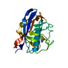
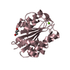
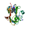
- PDB-5cu1: Crystal structure of DMSP lyase DddQ from Ruegeria pomeroyi DSS-3 -
+ Open data

- Basic information
| Entry | Database: PDB / ID: 5cu1 | ||||||
|---|---|---|---|---|---|---|---|
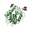
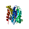
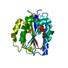
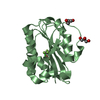
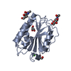
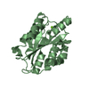
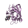
| Title | Crystal structure of DMSP lyase DddQ from Ruegeria pomeroyi DSS-3 | ||||||
 Components Components | DMSP lyase | ||||||
 Keywords Keywords | LYASE / Metalloenzyme / DMSP / Cupin | ||||||
| Function / homology |  Function and homology information Function and homology informationdimethylpropiothetin dethiomethylase / dimethylpropiothetin dethiomethylase activity / metal ion binding Similarity search - Function | ||||||
| Biological species |  Ruegeria pomeroyi (bacteria) Ruegeria pomeroyi (bacteria) | ||||||
| Method |  X-RAY DIFFRACTION / SYNCHROTRON / MOLECULAR REPLACEMENT / Resolution: 2.3 Å X-RAY DIFFRACTION / SYNCHROTRON / MOLECULAR REPLACEMENT / Resolution: 2.3 Å | ||||||
 Authors Authors | Chong, S.Y. / Moran, M.A. / Lanzilotta, W.N. / Whitman, W.B. | ||||||
 Citation Citation | Journal: To Be Published Title: To be Published Authors: Chong, S.Y. / Lanzilotta, W.N. | ||||||
| History |
|
- Structure visualization
| Structure viewer | Molecule: MolmilJmol/JSmol |
|---|
- Downloads & links
-Download
| PDBx/mmCIF format | 5cu1.cif.gz | 58 KB | Display | PDBx/mmCIF format |
|---|---|---|---|---|
| PDB format | pdb5cu1.ent.gz | 37.7 KB | Display | PDB format |
| PDBx/mmJSON format | 5cu1.json.gz | Tree view | PDBx/mmJSON format | |
| Others |  Other downloads Other downloads |
-Validation report
| Arichive directory | https://data.pdbj.org/pub/pdb/validation_reports/cu/5cu1ftp://data.pdbj.org/pub/pdb/validation_reports/cu/5cu1 | HTTPS FTP |
|---|
-Related structure data
| Related structure data |  4la2S S: Starting model for refinement |
|---|---|
| Similar structure data |
-Links
 PDBj
PDBj- Assembly
| Deposited unit | 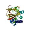
| ||||||||
|---|---|---|---|---|---|---|---|---|---|
| 1 |
| ||||||||
| Unit cell |
|
-Components
| #1: Protein | Mass: 23360.342 Da / Num. of mol.: 1 Source method: isolated from a genetically manipulated source Source: (gene. exp.) Ruegeria pomeroyi (strain ATCC 700808 / DSM 15171 / DSS-3) (bacteria)Strain: ATCC 700808 / DSM 15171 / DSS-3 / Gene: dddQ, SPO1596 / Production host: |
|---|---|
| #2: Chemical | ChemComp-FE /   Mass: 55.845 Da / Num. of mol.: 1 / Source method: obtained synthetically / Formula: Fe Mass: 55.845 Da / Num. of mol.: 1 / Source method: obtained synthetically / Formula: Fe |
| #3: Water | ChemComp-HOH /  Mass: 18.015 Da / Num. of mol.: 62 / Source method: isolated from a natural source / Formula: H2O Mass: 18.015 Da / Num. of mol.: 62 / Source method: isolated from a natural source / Formula: H2O |
-Experimental details
-Experiment
| Experiment | Method: X-RAY DIFFRACTION |
|---|
- Sample preparation
| Crystal | Density Matthews: 2.46 Å3/Da / Density % sol: 50.04 % |
|---|---|
| Crystal grow | Temperature: 291 K / Method: vapor diffusion, sitting drop / pH: 5.5 Details: 0.1M ammonium acetate, 0.1M bis-tris pH 5.5, 17% w/v PEG 10,000 |
-Data collection
| Diffraction | Mean temperature: 100 K |
|---|---|
| Diffraction source | Source: SYNCHROTRON / Site: APS  / Beamline: 22-BM / Wavelength: 1 Å / Beamline: 22-BM / Wavelength: 1 Å |
| Detector | Type: MARMOSAIC 225 mm CCD / Detector: CCD / Date: Oct 15, 2014 |
| Radiation | Monochromator: SI 111 crystal / Protocol: SINGLE WAVELENGTH / Monochromatic (M) / Laue (L): M / Scattering type: x-ray |
| Radiation wavelength | Wavelength: 1 Å / Relative weight: 1 |
| Reflection | Resolution: 2.3→46.62 Å / Num. obs: 10150 / % possible obs: 99.9 % / Redundancy: 5.9 % / Rsym value: 1.071 / Net I/σ(I): 9.69 |
| Reflection shell | Resolution: 2.3→2.38 Å / Redundancy: 4.7 % / Mean I/σ(I) obs: 1.95 / % possible all: 93.7 |
- Processing
| Software |
| ||||||||||||||||||||||||||||
|---|---|---|---|---|---|---|---|---|---|---|---|---|---|---|---|---|---|---|---|---|---|---|---|---|---|---|---|---|---|
| Refinement | Method to determine structure: MOLECULAR REPLACEMENT Starting model: 4LA2 Resolution: 2.3→46.619 Å / SU ML: 0.26 / Cross valid method: FREE R-VALUE / σ(F): 1.35 / Phase error: 28.88 / Stereochemistry target values: ML
| ||||||||||||||||||||||||||||
| Solvent computation | Shrinkage radii: 0.9 Å / VDW probe radii: 1.11 Å / Solvent model: FLAT BULK SOLVENT MODEL | ||||||||||||||||||||||||||||
| Refinement step | Cycle: LAST / Resolution: 2.3→46.619 Å
| ||||||||||||||||||||||||||||
| Refine LS restraints |
| ||||||||||||||||||||||||||||
| LS refinement shell |
|
