 Movie
Movie Controller


+ Open data

- Basic information
| Entry | Database: PDB / ID: 1h0p | ||||||
|---|---|---|---|---|---|---|---|
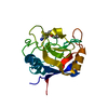
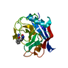
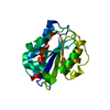
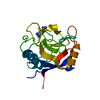
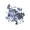
| Title | Cyclophilin_5 from C. elegans | ||||||
 Components Components | PEPTIDYL-PROLYL CIS-TRANS ISOMERASE 5 | ||||||
 Keywords Keywords | ISOMERASE / ROTAMASE | ||||||
| Function / homology |  Function and homology information Function and homology informationcyclosporin A binding / peptidylprolyl isomerase / peptidyl-prolyl cis-trans isomerase activity / protein folding / cytoplasm Similarity search - Function | ||||||
| Biological species |  | ||||||
| Method |  X-RAY DIFFRACTION / SYNCHROTRON / MOLECULAR REPLACEMENT / Resolution: 1.75 Å X-RAY DIFFRACTION / SYNCHROTRON / MOLECULAR REPLACEMENT / Resolution: 1.75 Å | ||||||
 Authors Authors | Picken, N.C. / Eschenlauer, S. / Taylor, P. / Page, A.P. / Walkinshaw, M.D. | ||||||
 Citation Citation | Journal: J.Mol.Biol. / Year: 2002 Title: Structural and Biological Characterisation of the Gut-Associated Cyclophilin B Isoforms from Caenorhabditis Elegans Authors: Picken, N.C. / Eschenlauer, S. / Taylor, P. / Page, A.P. / Walkinshaw, M.D. | ||||||
| History |
|
- Structure visualization
| Structure viewer | Molecule: MolmilJmol/JSmol |
|---|
- Downloads & links
-Download
| PDBx/mmCIF format | 1h0p.cif.gz | 53.3 KB | Display | PDBx/mmCIF format |
|---|---|---|---|---|
| PDB format | pdb1h0p.ent.gz | 36.8 KB | Display | PDB format |
| PDBx/mmJSON format | 1h0p.json.gz | Tree view | PDBx/mmJSON format | |
| Others |  Other downloads Other downloads |
-Validation report
| Arichive directory | https://data.pdbj.org/pub/pdb/validation_reports/h0/1h0pftp://data.pdbj.org/pub/pdb/validation_reports/h0/1h0p | HTTPS FTP |
|---|
-Related structure data
| Related structure data | 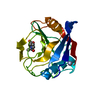 1e8kS S: Starting model for refinement |
|---|---|
| Similar structure data |


-Links
 PDBj
PDBj


- Assembly
| Deposited unit | 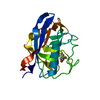
| ||||||||
|---|---|---|---|---|---|---|---|---|---|
| 1 |
| ||||||||
| Unit cell |
|
-Components
| #1: Protein | Mass: 19890.775 Da / Num. of mol.: 1 / Fragment: RESIDUES 23-204 / Mutation: YES Source method: isolated from a genetically manipulated source Source: (gene. exp.)  |
|---|---|
| #2: Chemical | ChemComp-DTT /   Mass: 154.251 Da / Num. of mol.: 1 / Source method: obtained synthetically / Formula: C4H10O2S2 Mass: 154.251 Da / Num. of mol.: 1 / Source method: obtained synthetically / Formula: C4H10O2S2 |
| #3: Water | ChemComp-HOH /  Mass: 18.015 Da / Num. of mol.: 201 / Source method: isolated from a natural source / Formula: H2O Mass: 18.015 Da / Num. of mol.: 201 / Source method: isolated from a natural source / Formula: H2O |
-Experimental details
-Experiment
| Experiment | Method: X-RAY DIFFRACTION / Number of used crystals: 1 |
|---|
- Sample preparation
| Crystal | Density Matthews: 2.4 Å3/Da / Density % sol: 47.9 % | ||||||||||||||||||||||||||||||||||||||||||||||||||||||||
|---|---|---|---|---|---|---|---|---|---|---|---|---|---|---|---|---|---|---|---|---|---|---|---|---|---|---|---|---|---|---|---|---|---|---|---|---|---|---|---|---|---|---|---|---|---|---|---|---|---|---|---|---|---|---|---|---|---|
| Crystal grow | pH: 6 / Details: PH 6.0, PEG 4000, LITHIUM CHLORIDE | ||||||||||||||||||||||||||||||||||||||||||||||||||||||||
| Crystal grow | *PLUS Temperature: 18 ℃ / Method: vapor diffusion, hanging drop | ||||||||||||||||||||||||||||||||||||||||||||||||||||||||
| Components of the solutions | *PLUS
|
-Data collection
| Diffraction | Mean temperature: 100 K |
|---|---|
| Diffraction source | Source: SYNCHROTRON / Site: SRS  / Beamline: PX14.1 / Wavelength: 1.488 / Beamline: PX14.1 / Wavelength: 1.488 |
| Detector | Type: ADSC CCD / Detector: CCD |
| Radiation | Protocol: SINGLE WAVELENGTH / Monochromatic (M) / Laue (L): M / Scattering type: x-ray |
| Radiation wavelength | Wavelength: 1.488 Å / Relative weight: 1 |
| Reflection | Resolution: 1.75→24 Å / Num. obs: 21525 / % possible obs: 99.9 % / Observed criterion σ(I): 3 / Redundancy: 9.5 % / Rmerge(I) obs: 0.086 / Net I/σ(I): 27.7 |
| Reflection shell | Resolution: 1.75→1.78 Å / Redundancy: 7.5 % / Rmerge(I) obs: 0.53 / Mean I/σ(I) obs: 3.7 / % possible all: 100 |
| Reflection | *PLUS Lowest resolution: 23.5 Å |
| Reflection shell | *PLUS % possible obs: 100 % |
- Processing
| Software |
| |||||||||||||||||||||||||||||||||
|---|---|---|---|---|---|---|---|---|---|---|---|---|---|---|---|---|---|---|---|---|---|---|---|---|---|---|---|---|---|---|---|---|---|---|
| Refinement | Method to determine structure: MOLECULAR REPLACEMENT Starting model: PDB ENTRY 1E8K Resolution: 1.75→10 Å / Num. parameters: 6488 / Num. restraintsaints: 5794 / Cross valid method: FREE R-VALUE / σ(F): 0 / Stereochemistry target values: ENGH AND HUBER
| |||||||||||||||||||||||||||||||||
| Solvent computation | Solvent model: MOEWS & KRETSINGER | |||||||||||||||||||||||||||||||||
| Refine analyze | Num. disordered residues: 2 / Occupancy sum non hydrogen: 1608.3 | |||||||||||||||||||||||||||||||||
| Refinement step | Cycle: LAST / Resolution: 1.75→10 Å
| |||||||||||||||||||||||||||||||||
| Refine LS restraints |
| |||||||||||||||||||||||||||||||||
| Software | *PLUS Name: SHELXL / Version: 97 / Classification: refinement | |||||||||||||||||||||||||||||||||
| Refinement | *PLUS Lowest resolution: 23.5 Å / % reflection Rfree: 5 % / Rfactor all: 0.18 / Rfactor obs: 0.236 / Rfactor Rfree: 0.169 | |||||||||||||||||||||||||||||||||
| Solvent computation | *PLUS | |||||||||||||||||||||||||||||||||
| Displacement parameters | *PLUS | |||||||||||||||||||||||||||||||||
| Refine LS restraints | *PLUS
|
