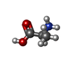
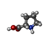
+ Open data
- Basic information
| Entry | Database: PDB / ID: 1e8k | ||||||
|---|---|---|---|---|---|---|---|
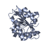
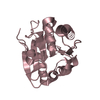
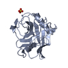
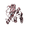
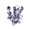
| Title | Cyclophilin 3 Complexed With Dipeptide Ala-Pro | ||||||
 Components Components | PEPTIDYL-PROLYL CIS-TRANS ISOMERASE 3 | ||||||
 Keywords Keywords | ISOMERASE | ||||||
| Function / homology |  Function and homology information Function and homology informationcyclosporin A binding / peptidylprolyl isomerase / peptidyl-prolyl cis-trans isomerase activity / protein folding / cytoplasm Similarity search - Function | ||||||
| Biological species |  | ||||||
| Method |  X-RAY DIFFRACTION / X-RAY DIFFRACTION /  MOLECULAR REPLACEMENT / Resolution: 1.9 Å MOLECULAR REPLACEMENT / Resolution: 1.9 Å | ||||||
 Authors Authors | Wu, S.Y. / Dornan, J. / Kontopidis, G. / Taylor, P. / Walkinshaw, M.D. | ||||||
 Citation Citation |  Journal: Angew.Chem.Int.Ed.Engl. / Year: 2001 Journal: Angew.Chem.Int.Ed.Engl. / Year: 2001Title: The First Direct Determination of a Ligand Binding Constant in Protein Crystals Authors: Wu Sy, S.Y. / Dornan, J. / Kontopidis, G. / Taylor, P. / Walkinshaw, M.D. #1:  Journal: J.Biol.Chem. / Year: 1999 Journal: J.Biol.Chem. / Year: 1999Title: Biochemical and Structural Characterization Ivergent Loop Cyclophilin from Caenorhabditis Elegans of A Authors: Dornan, J. / Page, A.P. / Taylor, P. / Wu, S.Y. / Winter, A.D. / Husi, H. / Walkinshawr, M.D. | ||||||
| History |
|
- Structure visualization
| Structure viewer | Molecule:  Molmil Molmil Jmol/JSmol Jmol/JSmol |
|---|
- Downloads & links
- Download
| PDBx/mmCIF format |  1e8k.cif.gz 1e8k.cif.gz | 51.9 KB | Display |  PDBx/mmCIF format PDBx/mmCIF format |
|---|---|---|---|---|
| PDB format |  pdb1e8k.ent.gz pdb1e8k.ent.gz | 36.1 KB | Display |  PDB format PDB format |
| PDBx/mmJSON format |  1e8k.json.gz 1e8k.json.gz | Tree view |  PDBx/mmJSON format PDBx/mmJSON format | |
| Others |  Other downloads Other downloads |
-Validation report
| Summary document |  1e8k_validation.pdf.gz 1e8k_validation.pdf.gz | 390.7 KB | Display |  wwPDB validaton report wwPDB validaton report |
|---|---|---|---|---|
| Full document |  1e8k_full_validation.pdf.gz 1e8k_full_validation.pdf.gz | 393.3 KB | Display | |
| Data in XML |  1e8k_validation.xml.gz 1e8k_validation.xml.gz | 5.3 KB | Display | |
| Data in CIF |  1e8k_validation.cif.gz 1e8k_validation.cif.gz | 8.8 KB | Display | |
| Arichive directory |  https://data.pdbj.org/pub/pdb/validation_reports/e8/1e8k https://data.pdbj.org/pub/pdb/validation_reports/e8/1e8k ftp://data.pdbj.org/pub/pdb/validation_reports/e8/1e8k ftp://data.pdbj.org/pub/pdb/validation_reports/e8/1e8k | HTTPS FTP |
-Related structure data
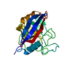
| Related structure data | 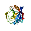 1dywS S: Starting model for refinement |
|---|---|
| Similar structure data |
- Links
Links
- Assembly
| Deposited unit | 
| ||||||||
|---|---|---|---|---|---|---|---|---|---|
| 1 |
| ||||||||
| Unit cell |
| ||||||||
| Components on special symmetry positions |
|
- Components
| #1: Protein | Mass: 18576.182 Da / Num. of mol.: 1 Source method: isolated from a genetically manipulated source Source: (gene. exp.)   |
|---|---|
| #2: Chemical | ChemComp-ALA / |
| #3: Chemical | ChemComp-PRO / |
| #4: Water | ChemComp-HOH / |
-Experimental details
-Experiment
| Experiment | Method:  X-RAY DIFFRACTION / Number of used crystals: 1 X-RAY DIFFRACTION / Number of used crystals: 1 |
|---|
- Sample preparation
| Crystal | Density Matthews: 3.08 Å3/Da / Density % sol: 59.73 % | ||||||||||||||||||||||||||||||
|---|---|---|---|---|---|---|---|---|---|---|---|---|---|---|---|---|---|---|---|---|---|---|---|---|---|---|---|---|---|---|---|
| Crystal grow | pH: 5.6 / Details: MPEG5000, SODIUM CITRATE, PH 5.6 | ||||||||||||||||||||||||||||||
| Crystal grow | *PLUS Temperature: 16 ℃ / Method: vapor diffusion, hanging drop / Details: Dornan, J., (1999) J.Biol.Chem., 274, 34877. | ||||||||||||||||||||||||||||||
| Components of the solutions | *PLUS
|
-Data collection
| Diffraction | Mean temperature: 100 K |
|---|---|
| Diffraction source | Source:  ROTATING ANODE / Type: ENRAF-NONIUS FR571 / Wavelength: 1.5418 ROTATING ANODE / Type: ENRAF-NONIUS FR571 / Wavelength: 1.5418 |
| Detector | Type: MARRESEARCH / Detector: IMAGE PLATE / Details: COLLIMATOR |
| Radiation | Monochromator: GRAPHITE / Protocol: SINGLE WAVELENGTH / Monochromatic (M) / Laue (L): M / Scattering type: x-ray |
| Radiation wavelength | Wavelength: 1.5418 Å / Relative weight: 1 |
| Reflection | Resolution: 1.9→25 Å / Num. obs: 19230 / % possible obs: 97.6 % / Redundancy: 7.92 % / Rsym value: 0.079 / Net I/σ(I): 12.85 |
| Reflection shell | Resolution: 1.9→1.93 Å / Mean I/σ(I) obs: 2.13 / Rsym value: 0.332 / % possible all: 88.2 |
| Reflection | *PLUS Num. measured all: 152448 / Rmerge(I) obs: 0.079 |
| Reflection shell | *PLUS Rmerge(I) obs: 0.332 |
- Processing
| Software |
| |||||||||||||||||||||||||||||||||
|---|---|---|---|---|---|---|---|---|---|---|---|---|---|---|---|---|---|---|---|---|---|---|---|---|---|---|---|---|---|---|---|---|---|---|
| Refinement | Method to determine structure:  MOLECULAR REPLACEMENT MOLECULAR REPLACEMENTStarting model: PDB ENTRY 1DYW Resolution: 1.9→10 Å / Num. parameters: 6247 / Num. restraintsaints: 5392 / Cross valid method: FREE R-VALUE / σ(F): 0 / Stereochemistry target values: ENGH AND HUBER
| |||||||||||||||||||||||||||||||||
| Solvent computation | Solvent model: MOEWS & KRETSINGER, J.MOL.BIOL.91(1973)201-2 | |||||||||||||||||||||||||||||||||
| Refine analyze | Num. disordered residues: 0 / Occupancy sum hydrogen: 0 / Occupancy sum non hydrogen: 1497.31 | |||||||||||||||||||||||||||||||||
| Refinement step | Cycle: LAST / Resolution: 1.9→10 Å
| |||||||||||||||||||||||||||||||||
| Refine LS restraints |
| |||||||||||||||||||||||||||||||||
| Software | *PLUS Name: SHELXL-97 / Classification: refinement | |||||||||||||||||||||||||||||||||
| Refinement | *PLUS % reflection Rfree: 5 % / Rfactor Rwork: 0.1826 | |||||||||||||||||||||||||||||||||
| Solvent computation | *PLUS | |||||||||||||||||||||||||||||||||
| Displacement parameters | *PLUS | |||||||||||||||||||||||||||||||||
| Refine LS restraints | *PLUS Type: s_bond_d / Dev ideal: 0.03 |
 Movie
Movie Controller






 PDBj
PDBj


