 Movie
Movie Controller

[English] 日本語
 Yorodumi
- PDB-3d2b: Structure of 2D9, a thermostable mutant of Bacillus subtilis lipa... -
+ Open data

- Basic information
| Entry | Database: PDB / ID: 3d2b | ||||||
|---|---|---|---|---|---|---|---|
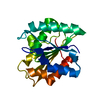
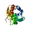
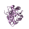
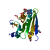
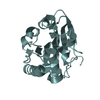
| Title | Structure of 2D9, a thermostable mutant of Bacillus subtilis lipase obtained through directed evolution | ||||||
 Components Components | Lipase | ||||||
 Keywords Keywords | HYDROLASE / lipase / alpha/beta hydrolase / stability / directed evolution / Lipid degradation / Secreted | ||||||
| Function / homology |  Function and homology information Function and homology informationlipase activity / triacylglycerol lipase / triacylglycerol lipase activity / lipid catabolic process / extracellular region Similarity search - Function | ||||||
| Biological species |  | ||||||
| Method |  X-RAY DIFFRACTION / MOLECULAR REPLACEMENT / molecular replacement / Resolution: 1.95 Å X-RAY DIFFRACTION / MOLECULAR REPLACEMENT / molecular replacement / Resolution: 1.95 Å | ||||||
 Authors Authors | Sankaranarayanan, R. / Kamal, M.Z. | ||||||
 Citation Citation | Journal: J.Mol.Biol. / Year: 2008 Title: Thermostable Bacillus subtilis lipases: in vitro evolution and structural insight Authors: Ahmad, S. / Kamal, M.Z. / Sankaranarayanan, R. / Rao, N.M. | ||||||
| History |
|
- Structure visualization
| Structure viewer | Molecule: MolmilJmol/JSmol |
|---|
- Downloads & links
-Download
| PDBx/mmCIF format | 3d2b.cif.gz | 83.6 KB | Display | PDBx/mmCIF format |
|---|---|---|---|---|
| PDB format | pdb3d2b.ent.gz | 63.7 KB | Display | PDB format |
| PDBx/mmJSON format | 3d2b.json.gz | Tree view | PDBx/mmJSON format | |
| Others |  Other downloads Other downloads |
-Validation report
| Arichive directory | https://data.pdbj.org/pub/pdb/validation_reports/d2/3d2bftp://data.pdbj.org/pub/pdb/validation_reports/d2/3d2b | HTTPS FTP |
|---|
-Related structure data


-Links
 PDBj
PDBj- Assembly
| Deposited unit | 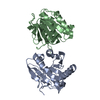
| ||||||||
|---|---|---|---|---|---|---|---|---|---|
| 1 | 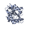
| ||||||||
| 2 | 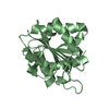
| ||||||||
| Unit cell |
|
-Components
| #1: Protein | Mass: 19466.889 Da / Num. of mol.: 2 / Mutation: F17S, N89Y, L114P, A132D, I157M, N166Y Source method: isolated from a genetically manipulated source Source: (gene. exp.) #2: Water | ChemComp-HOH / |  Mass: 18.015 Da / Num. of mol.: 313 / Source method: isolated from a natural source / Formula: H2O Mass: 18.015 Da / Num. of mol.: 313 / Source method: isolated from a natural source / Formula: H2O |
|---|
-Experimental details
-Experiment
| Experiment | Method: X-RAY DIFFRACTION / Number of used crystals: 1 |
|---|
- Sample preparation
| Crystal | Density Matthews: 3.33 Å3/Da / Density % sol: 63.09 % / Mosaicity: 0.593 ° |
|---|---|
| Crystal grow | Temperature: 277 K / Method: vapor diffusion, sitting drop / pH: 9.5 Details: 100mM Ethanolamine buffe, 10mM Sodium sulfate, pH 9.5, vapour diffusion, sitting drop, temperature 277K, VAPOR DIFFUSION, SITTING DROP |
-Data collection
| Diffraction | Mean temperature: 100 K | ||||||||||||||||||||||||||||||||||||||||||||||||||||||||||||||||||
|---|---|---|---|---|---|---|---|---|---|---|---|---|---|---|---|---|---|---|---|---|---|---|---|---|---|---|---|---|---|---|---|---|---|---|---|---|---|---|---|---|---|---|---|---|---|---|---|---|---|---|---|---|---|---|---|---|---|---|---|---|---|---|---|---|---|---|---|
| Diffraction source | Source: ROTATING ANODE / Type: RIGAKU RUH3R / Wavelength: 1.5418 Å | ||||||||||||||||||||||||||||||||||||||||||||||||||||||||||||||||||
| Detector | Type: MAR scanner 345 mm plate / Detector: IMAGE PLATE / Details: osmic mirrors | ||||||||||||||||||||||||||||||||||||||||||||||||||||||||||||||||||
| Radiation | Protocol: SINGLE WAVELENGTH / Monochromatic (M) / Laue (L): M / Scattering type: x-ray | ||||||||||||||||||||||||||||||||||||||||||||||||||||||||||||||||||
| Radiation wavelength | Wavelength: 1.5418 Å / Relative weight: 1 | ||||||||||||||||||||||||||||||||||||||||||||||||||||||||||||||||||
| Reflection | Resolution: 1.95→25 Å / Num. obs: 36223 / % possible obs: 94.3 % / Redundancy: 3.6 % / Rmerge(I) obs: 0.066 / Χ2: 0.561 / Net I/σ(I): 10.2 | ||||||||||||||||||||||||||||||||||||||||||||||||||||||||||||||||||
| Reflection shell |
|
-Phasing
| Phasing | Method: molecular replacement |
|---|
- Processing
| Software |
| ||||||||||||||||||||||||
|---|---|---|---|---|---|---|---|---|---|---|---|---|---|---|---|---|---|---|---|---|---|---|---|---|---|
| Refinement | Method to determine structure: MOLECULAR REPLACEMENT / Resolution: 1.95→25 Å / FOM work R set: 0.885 / σ(F): 0
| ||||||||||||||||||||||||
| Solvent computation | Bsol: 41.541 Å2 | ||||||||||||||||||||||||
| Displacement parameters | Biso mean: 15.955 Å2
| ||||||||||||||||||||||||
| Refinement step | Cycle: LAST / Resolution: 1.95→25 Å
| ||||||||||||||||||||||||
| Refine LS restraints |
| ||||||||||||||||||||||||
| Xplor file |
|
