 Movie
Movie Controller


+ Open data

- Basic information
| Entry | Database: PDB / ID: 5mvr | ||||||
|---|---|---|---|---|---|---|---|
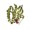
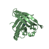
| Title | Crystal structure of Bacillus subtilus YdiB | ||||||
 Components Components | tRNA threonylcarbamoyladenosine biosynthesis protein TsaE | ||||||
 Keywords Keywords | TRANSFERASE / kinase / ADP / phosphorylation | ||||||
| Function / homology | tRNA threonylcarbamoyl adenosine modification protein TsaE / Threonylcarbamoyl adenosine biosynthesis protein TsaE / tRNA threonylcarbamoyladenosine modification / P-loop containing nucleoside triphosphate hydrolase / ATP binding / metal ion binding / cytoplasm / ADENOSINE-5'-DIPHOSPHATE / tRNA threonylcarbamoyladenosine biosynthesis protein TsaE Function and homology information Function and homology information | ||||||
| Biological species |  | ||||||
| Method |  X-RAY DIFFRACTION / SYNCHROTRON / MOLECULAR REPLACEMENT / Resolution: 1.762 Å X-RAY DIFFRACTION / SYNCHROTRON / MOLECULAR REPLACEMENT / Resolution: 1.762 Å | ||||||
 Authors Authors | Jault, J.-M. / Aghajari, N. | ||||||
 Citation Citation | Journal: J. Mol. Biol. / Year: 2017 Title: Expanding the Kinome World: A New Protein Kinase Family Widely Conserved in Bacteria. Authors: Nguyen, H.A. / El Khoury, T. / Guiral, S. / Laaberki, M.H. / Candusso, M.P. / Galisson, F. / Foucher, A.E. / Kesraoui, S. / Ballut, L. / Vallet, S. / Orelle, C. / Zucchini, L. / Martin, J. / ...Authors: Nguyen, H.A. / El Khoury, T. / Guiral, S. / Laaberki, M.H. / Candusso, M.P. / Galisson, F. / Foucher, A.E. / Kesraoui, S. / Ballut, L. / Vallet, S. / Orelle, C. / Zucchini, L. / Martin, J. / Page, A. / Attieh, J. / Aghajari, N. / Grangeasse, C. / Jault, J.M. | ||||||
| History |
|
- Structure visualization
| Structure viewer | Molecule: MolmilJmol/JSmol |
|---|
- Downloads & links
-Download
| PDBx/mmCIF format | 5mvr.cif.gz | 81.6 KB | Display | PDBx/mmCIF format |
|---|---|---|---|---|
| PDB format | pdb5mvr.ent.gz | 59.4 KB | Display | PDB format |
| PDBx/mmJSON format | 5mvr.json.gz | Tree view | PDBx/mmJSON format | |
| Others |  Other downloads Other downloads |
-Validation report
| Arichive directory | https://data.pdbj.org/pub/pdb/validation_reports/mv/5mvrftp://data.pdbj.org/pub/pdb/validation_reports/mv/5mvr | HTTPS FTP |
|---|
-Related structure data
| Related structure data | 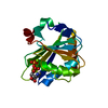 5np9C 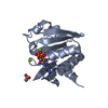 1htwS S: Starting model for refinement C: citing same article ( |
|---|---|
| Similar structure data |


-Links
 PDBj
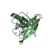
PDBj- Assembly
| Deposited unit | 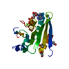
| ||||||||
|---|---|---|---|---|---|---|---|---|---|
| 1 |
| ||||||||
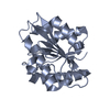
| Unit cell |
|
-Components
| #1: Protein | Mass: 18308.633 Da / Num. of mol.: 1 Source method: isolated from a genetically manipulated source Details: Chain break from residue 59-62 (incl.) and 85-90 (incl.) due to missing electron density. The three C-ter residues are lacking in the structure due to missing electron density. The four ...Details: Chain break from residue 59-62 (incl.) and 85-90 (incl.) due to missing electron density. The three C-ter residues are lacking in the structure due to missing electron density. The four initial N-ter residues come from the His-Tag. Mismatch is due to ADP aligning with C-ter residues Source: (gene. exp.) | ||
|---|---|---|---|
| #2: Chemical | ChemComp-ADP /   Mass: 427.201 Da / Num. of mol.: 1 / Source method: obtained synthetically / Formula: C10H15N5O10P2 / Comment: ADP, energy-carrying molecule*YM Mass: 427.201 Da / Num. of mol.: 1 / Source method: obtained synthetically / Formula: C10H15N5O10P2 / Comment: ADP, energy-carrying molecule*YM | ||
| #3: Chemical | ChemComp-MG /   Mass: 24.305 Da / Num. of mol.: 1 / Source method: isolated from a natural source / Formula: Mg Mass: 24.305 Da / Num. of mol.: 1 / Source method: isolated from a natural source / Formula: Mg | ||
| #4: Chemical |   Mass: 62.068 Da / Num. of mol.: 3 / Source method: obtained synthetically / Formula: C2H6O2 Mass: 62.068 Da / Num. of mol.: 3 / Source method: obtained synthetically / Formula: C2H6O2#5: Water | ChemComp-HOH / |  Mass: 18.015 Da / Num. of mol.: 57 / Source method: isolated from a natural source / Formula: H2O Mass: 18.015 Da / Num. of mol.: 57 / Source method: isolated from a natural source / Formula: H2O |
-Experimental details
-Experiment
| Experiment | Method: X-RAY DIFFRACTION / Number of used crystals: 1 |
|---|
- Sample preparation
| Crystal | Density Matthews: 2.07 Å3/Da / Density % sol: 40.58 % |
|---|---|
| Crystal grow | Temperature: 293 K / Method: vapor diffusion, sitting drop / Details: 42 % PEG600 and 200 mM Imidazole malate pH 5.5 |
-Data collection
| Diffraction | Mean temperature: 100 K |
|---|---|
| Diffraction source | Source: SYNCHROTRON / Site: ESRF  / Beamline: ID29 / Wavelength: 1.07252 Å / Beamline: ID29 / Wavelength: 1.07252 Å |
| Detector | Type: DECTRIS PILATUS 6M / Detector: PIXEL / Date: Sep 24, 2016 |
| Radiation | Monochromator: liquid nitrogen cooled channel-cut silicon monochromator Protocol: SINGLE WAVELENGTH / Monochromatic (M) / Laue (L): M / Scattering type: x-ray |
| Radiation wavelength | Wavelength: 1.07252 Å / Relative weight: 1 |
| Reflection | Resolution: 1.76→43.84 Å / Num. obs: 14655 / % possible obs: 98.6 % / Redundancy: 3.2 % / Biso Wilson estimate: 31.2 Å2 / Rmerge(I) obs: 0.032 / Net I/σ(I): 16.5 |
- Processing
| Software |
| ||||||||||||||||||||||||||||||||||||||||||
|---|---|---|---|---|---|---|---|---|---|---|---|---|---|---|---|---|---|---|---|---|---|---|---|---|---|---|---|---|---|---|---|---|---|---|---|---|---|---|---|---|---|---|---|
| Refinement | Method to determine structure: MOLECULAR REPLACEMENT Starting model: 1HTW Resolution: 1.762→43.835 Å / SU ML: 0.21 / Cross valid method: FREE R-VALUE / σ(F): 1.39 / Phase error: 27.01 / Stereochemistry target values: ML
| ||||||||||||||||||||||||||||||||||||||||||
| Solvent computation | Shrinkage radii: 0.9 Å / VDW probe radii: 1.11 Å / Solvent model: FLAT BULK SOLVENT MODEL | ||||||||||||||||||||||||||||||||||||||||||
| Refinement step | Cycle: LAST / Resolution: 1.762→43.835 Å
| ||||||||||||||||||||||||||||||||||||||||||
| Refine LS restraints |
| ||||||||||||||||||||||||||||||||||||||||||
| LS refinement shell |
| ||||||||||||||||||||||||||||||||||||||||||
| Refinement TLS params. | Method: refined / Origin x: -0.1199 Å / Origin y: 3.1225 Å / Origin z: 49.5559 Å
| ||||||||||||||||||||||||||||||||||||||||||
| Refinement TLS group | Selection details: all |
