 Movie
Movie Controller


+ Open data

- Basic information
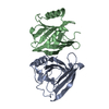
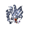
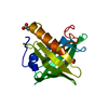
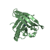
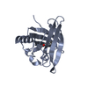
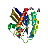
| Entry | Database: PDB / ID: 100 | ||||||
|---|---|---|---|---|---|---|---|
| Title | Porcine Odorant Binding Protein Complexed with undecanal | ||||||
 Components Components | ODORANT-BINDING PROTEIN | ||||||
 Keywords Keywords | ODORANT BINDING PROTEIN / LIPOCALINS | ||||||
| Function / homology |  Function and homology information Function and homology informationodorant binding / small molecule binding / sensory perception of smell / : Similarity search - Function | ||||||
| Biological species |  | ||||||
| Method |  X-RAY DIFFRACTION / SYNCHROTRON / OTHER / Resolution: 2.15 Å X-RAY DIFFRACTION / SYNCHROTRON / OTHER / Resolution: 2.15 Å | ||||||
 Authors Authors | Vincent, F. / Spinelli, S. / Cambillau, C. / Tegoni, M. | ||||||
 Citation Citation | Journal: J.Mol.Biol. / Year: 2000 Title: Complexes of Porcine Odorant Binding Protein with Odorant Molecules Belonging to Different Chemical Classes Authors: Vincent, F. / Spinelli, S. / Ramoni, R. / Grolli, S. / Pelosi, P. / Cambillau, C. / Tegoni, M. | ||||||
| History |
|
- Structure visualization
| Structure viewer | Molecule: MolmilJmol/JSmol |
|---|
- Downloads & links
-Download
| PDBx/mmCIF format | 1e02.cif.gz | 76.6 KB | Display | PDBx/mmCIF format |
|---|---|---|---|---|
| PDB format | pdb1e02.ent.gz | 57.7 KB | Display | PDB format |
| PDBx/mmJSON format | 1e02.json.gz | Tree view | PDBx/mmJSON format | |
| Others |  Other downloads Other downloads |
-Validation report
| Arichive directory | https://data.pdbj.org/pub/pdb/validation_reports/e0/1e02ftp://data.pdbj.org/pub/pdb/validation_reports/e0/1e02 | HTTPS FTP |
|---|
-Related structure data
| Related structure data | 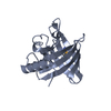 1dzjC  1dzkC  1dzmC  1dzpC 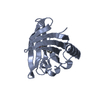 1e00C 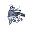 1e06C C: citing same article ( |
|---|---|
| Similar structure data |


-Links
 PDBj
PDBj

- Assembly
| Deposited unit | 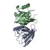
| ||||||||
|---|---|---|---|---|---|---|---|---|---|
| 1 | 
| ||||||||
| 2 | 
| ||||||||
| Unit cell |
| ||||||||
| Noncrystallographic symmetry (NCS) | NCS oper: (Code: given Matrix: (0.16058, -0.18129, 0.97023), Vector: |
-Components
| #1: Protein | Mass: 17721.414 Da / Num. of mol.: 2 / Source method: isolated from a natural source / Source: (natural) #2: Chemical |   Mass: 170.292 Da / Num. of mol.: 2 / Source method: obtained synthetically / Formula: C11H22O Mass: 170.292 Da / Num. of mol.: 2 / Source method: obtained synthetically / Formula: C11H22O#3: Water | ChemComp-HOH / |  Mass: 18.015 Da / Num. of mol.: 202 / Source method: isolated from a natural source / Formula: H2O Mass: 18.015 Da / Num. of mol.: 202 / Source method: isolated from a natural source / Formula: H2OHas protein modification | Y | |
|---|
-Experimental details
-Experiment
| Experiment | Method: X-RAY DIFFRACTION / Number of used crystals: 1 |
|---|
- Sample preparation
| Crystal | Density Matthews: 2.46 Å3/Da / Density % sol: 50 % | ||||||||||||||||||||
|---|---|---|---|---|---|---|---|---|---|---|---|---|---|---|---|---|---|---|---|---|---|
| Crystal grow | pH: 7.8 / Details: 2M AMMONIUM SULFATE, AND 5% ISOPROPANOL, pH 7.80 | ||||||||||||||||||||
| Crystal grow | *PLUS Temperature: 20 ℃ / Method: vapor diffusion | ||||||||||||||||||||
| Components of the solutions | *PLUS
|
-Data collection
| Diffraction | Mean temperature: 100 K |
|---|---|
| Diffraction source | Source: SYNCHROTRON / Site: LURE  / Beamline: DW32 / Wavelength: 0.97 / Beamline: DW32 / Wavelength: 0.97 |
| Detector | Type: MARRESEARCH / Detector: IMAGE PLATE / Date: Dec 1, 1997 |
| Radiation | Protocol: SINGLE WAVELENGTH / Monochromatic (M) / Laue (L): M / Scattering type: x-ray |
| Radiation wavelength | Wavelength: 0.97 Å / Relative weight: 1 |
| Reflection | Resolution: 1.83→15 Å / Num. obs: 30761 / % possible obs: 97.7 % / Redundancy: 2.9 % / Biso Wilson estimate: 13.5 Å2 / Rsym value: 0.047 / Net I/σ(I): 16 |
| Reflection shell | Resolution: 1.83→1.86 Å / Redundancy: 2.7 % / Mean I/σ(I) obs: 4.3 / Rsym value: 0.17 / % possible all: 96.9 |
| Reflection | *PLUS Highest resolution: 2.15 Å / Lowest resolution: 18 Å / % possible obs: 90.7 % / Redundancy: 3.2 % / Rmerge(I) obs: 0.079 |
| Reflection shell | *PLUS |
- Processing
| Software |
| ||||||||||||||||||||||||||||||||||||||||||||||||||||||||||||||||||||||||||||||||
|---|---|---|---|---|---|---|---|---|---|---|---|---|---|---|---|---|---|---|---|---|---|---|---|---|---|---|---|---|---|---|---|---|---|---|---|---|---|---|---|---|---|---|---|---|---|---|---|---|---|---|---|---|---|---|---|---|---|---|---|---|---|---|---|---|---|---|---|---|---|---|---|---|---|---|---|---|---|---|---|---|---|
| Refinement | Method to determine structure: OTHER / Resolution: 2.15→18 Å / Data cutoff high absF: 1000000 / Data cutoff low absF: 100 / Cross valid method: THROUGHOUT / σ(F): 0 Details: N-TERMINUS FROM RESIDUE 1 TO 8, IN SUBUNIT A AND 1 - 10 IN SUBUNIT B, ARE NOT VISIBLE IN THE ELECTRON DENSITY, DUE TO FLEXIBILITY. ALTERNATE POSITIONS ARE PRESENT FOR SIDE CHAIN OF RESIDUES ...Details: N-TERMINUS FROM RESIDUE 1 TO 8, IN SUBUNIT A AND 1 - 10 IN SUBUNIT B, ARE NOT VISIBLE IN THE ELECTRON DENSITY, DUE TO FLEXIBILITY. ALTERNATE POSITIONS ARE PRESENT FOR SIDE CHAIN OF RESIDUES 19A, 39A, 114A. THE ATOMS CONCERNED HAVE OCCUPANCY BETWEEN 0.0 AND 1.0 AND A SEGID AC1 AND AC2 OFTEN, OCCUPANCY VALUES LOWER THAN 1.0 APPEARED TO JUSTIFY BETTER THE ELECTRON DENSITY. FOR THIS REASON WE HAVE KEPT THIS LOW OCCUPANCY FOR SEVERAL SIDE CHAIN ATOMS.
| ||||||||||||||||||||||||||||||||||||||||||||||||||||||||||||||||||||||||||||||||
| Displacement parameters | Biso mean: 26.8 Å2
| ||||||||||||||||||||||||||||||||||||||||||||||||||||||||||||||||||||||||||||||||
| Refine analyze | Luzzati coordinate error obs: 0.32 Å / Luzzati d res low obs: 5 Å / Luzzati sigma a obs: 0.37 Å | ||||||||||||||||||||||||||||||||||||||||||||||||||||||||||||||||||||||||||||||||
| Refinement step | Cycle: LAST / Resolution: 2.15→18 Å
| ||||||||||||||||||||||||||||||||||||||||||||||||||||||||||||||||||||||||||||||||
| Refine LS restraints |
| ||||||||||||||||||||||||||||||||||||||||||||||||||||||||||||||||||||||||||||||||
| LS refinement shell | Resolution: 2.15→2.2 Å / Total num. of bins used: 15
| ||||||||||||||||||||||||||||||||||||||||||||||||||||||||||||||||||||||||||||||||
| Xplor file |
| ||||||||||||||||||||||||||||||||||||||||||||||||||||||||||||||||||||||||||||||||
| Software | *PLUS Name: X-PLOR / Version: 3.843 / Classification: refinement | ||||||||||||||||||||||||||||||||||||||||||||||||||||||||||||||||||||||||||||||||
| Refine LS restraints | *PLUS
|
