 Movie
Movie Controller


+ Open data

- Basic information
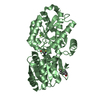
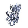
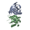
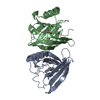
| Entry | Database: PDB / ID: 1a3y | ||||||
|---|---|---|---|---|---|---|---|
| Title | ODORANT BINDING PROTEIN FROM NASAL MUCOSA OF PIG | ||||||
 Components Components | ODORANT BINDING PROTEIN | ||||||
 Keywords Keywords | LIPOCALIN / OLFACTION | ||||||
| Function / homology |  Function and homology information Function and homology informationodorant binding / small molecule binding / sensory perception of smell / : Similarity search - Function | ||||||
| Biological species |  | ||||||
| Method |  X-RAY DIFFRACTION / MOLECULAR REPLACEMENT / Resolution: 2.25 Å X-RAY DIFFRACTION / MOLECULAR REPLACEMENT / Resolution: 2.25 Å | ||||||
 Authors Authors | Spinelli, S. / Cambillau, C. / Tegoni, M. | ||||||
 Citation Citation | Journal: Biochemistry / Year: 1998 Title: The structure of the monomeric porcine odorant binding protein sheds light on the domain swapping mechanism. Authors: Spinelli, S. / Ramoni, R. / Grolli, S. / Bonicel, J. / Cambillau, C. / Tegoni, M. #1: Journal: Chem.Senses / Year: 1995Title: Affinities of Nutty and Green-Smelling Pyrazines and Thiazoles to Odorant-Binding Proteins, in Relation with Their Lipophilicity Authors: Herent, M.F. / Collin, S. / Pelosi, P. #2: Journal: Chem.Senses / Year: 1993Title: Binding of Selected Odorant to Bovine and Porcine Odorant-Binding Proteins Authors: Monte, M.D. / Centini, M. / Anselmi, C. / Pelosi, P. #3: Journal: COMP.BIOCHEM.PHYSIOL. B: BIOCHEM.MOL.BIOL. / Year: 1991Title: Purification and Characterization of Two Odorant-Binding Proteins from Nasal Tissue of Rabbit and Pig Authors: Dal Monte, M. / Andreini, I. / Revoltella, R. / Pelosi, P. | ||||||
| History |
|
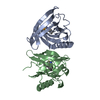
- Structure visualization
| Structure viewer | Molecule: MolmilJmol/JSmol |
|---|
- Downloads & links
-Download
| PDBx/mmCIF format | 1a3y.cif.gz | 89.9 KB | Display | PDBx/mmCIF format |
|---|---|---|---|---|
| PDB format | pdb1a3y.ent.gz | 69.3 KB | Display | PDB format |
| PDBx/mmJSON format | 1a3y.json.gz | Tree view | PDBx/mmJSON format | |
| Others |  Other downloads Other downloads |
-Validation report
| Arichive directory | https://data.pdbj.org/pub/pdb/validation_reports/a3/1a3yftp://data.pdbj.org/pub/pdb/validation_reports/a3/1a3y | HTTPS FTP |
|---|
-Related structure data
| Related structure data |  1obpS S: Starting model for refinement |
|---|---|
| Similar structure data |


-Links
 PDBj
PDBj

- Assembly
| Deposited unit | 
| ||||||||
|---|---|---|---|---|---|---|---|---|---|
| 1 |
| ||||||||
| Unit cell |
|
-Components
| #1: Protein | Mass: 16769.480 Da / Num. of mol.: 2 / Source method: isolated from a natural source / Source: (natural) #2: Water | ChemComp-HOH / |  Mass: 18.015 Da / Num. of mol.: 187 / Source method: isolated from a natural source / Formula: H2O Mass: 18.015 Da / Num. of mol.: 187 / Source method: isolated from a natural source / Formula: H2OHas protein modification | Y | |
|---|
-Experimental details
-Experiment
| Experiment | Method: X-RAY DIFFRACTION / Number of used crystals: 1 |
|---|
- Sample preparation
| Crystal | Density Matthews: 2.3 Å3/Da / Density % sol: 46 % | ||||||||||||||||||||
|---|---|---|---|---|---|---|---|---|---|---|---|---|---|---|---|---|---|---|---|---|---|
| Crystal grow | pH: 5.5 / Details: pH 5.5 | ||||||||||||||||||||
| Crystal grow | *PLUS Temperature: 20 ℃ / Method: vapor diffusion | ||||||||||||||||||||
| Components of the solutions | *PLUS
|
-Data collection
| Diffraction | Mean temperature: 100 K |
|---|---|
| Diffraction source | Source: ROTATING ANODE / Type: RIGAKU RUH2R / Wavelength: 1.5418 |
| Detector | Type: MARRESEARCH / Detector: IMAGE PLATE / Date: Jun 1, 1997 |
| Radiation | Monochromator: GRAPHITE(002) / Monochromatic (M) / Laue (L): M / Scattering type: x-ray |
| Radiation wavelength | Wavelength: 1.5418 Å / Relative weight: 1 |
| Reflection | Resolution: 2.15→30 Å / Num. obs: 11000 / % possible obs: 99.4 % / Observed criterion σ(I): 2 / Redundancy: 5 % / Rsym value: 0.051 / Net I/σ(I): 12 |
| Reflection shell | Resolution: 2.15→2.2 Å / Redundancy: 5 % / Mean I/σ(I) obs: 2 / Rsym value: 0.265 / % possible all: 99 |
| Reflection | *PLUS Rmerge(I) obs: 0.051 |
| Reflection shell | *PLUS % possible obs: 99 % / Rmerge(I) obs: 0.265 |
- Processing
| Software |
| ||||||||||||||||||||||||||||||||||||||||||||||||||||||||||||
|---|---|---|---|---|---|---|---|---|---|---|---|---|---|---|---|---|---|---|---|---|---|---|---|---|---|---|---|---|---|---|---|---|---|---|---|---|---|---|---|---|---|---|---|---|---|---|---|---|---|---|---|---|---|---|---|---|---|---|---|---|---|
| Refinement | Method to determine structure: MOLECULAR REPLACEMENT Starting model: 1OBP Resolution: 2.25→25 Å / Rfactor Rfree error: 0.0002 / Data cutoff high absF: 10000000 / Data cutoff low absF: 100 / Cross valid method: THROUGHOUT / σ(F): 0
| ||||||||||||||||||||||||||||||||||||||||||||||||||||||||||||
| Refinement step | Cycle: LAST / Resolution: 2.25→25 Å
| ||||||||||||||||||||||||||||||||||||||||||||||||||||||||||||
| Refine LS restraints |
| ||||||||||||||||||||||||||||||||||||||||||||||||||||||||||||
| LS refinement shell | Resolution: 2.25→2.37 Å / Rfactor Rfree error: 0.011 / Total num. of bins used: 7
| ||||||||||||||||||||||||||||||||||||||||||||||||||||||||||||
| Xplor file |
| ||||||||||||||||||||||||||||||||||||||||||||||||||||||||||||
| Software | *PLUS Name: X-PLOR / Version: 3.843 / Classification: refinement | ||||||||||||||||||||||||||||||||||||||||||||||||||||||||||||
| Refine LS restraints | *PLUS
|
