 Movie
Movie Controller


+ Open data

- Basic information
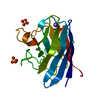
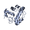
| Entry | Database: PDB / ID: 5np9 | ||||||
|---|---|---|---|---|---|---|---|
| Title | Crystal structure of Bacillus subtilis YdiB in complex with ADP | ||||||
 Components Components | tRNA threonylcarbamoyladenosine biosynthesis protein TsaE | ||||||
 Keywords Keywords | TRANSFERASE / Kinase / ADP / phosphorylastion | ||||||
| Function / homology | tRNA threonylcarbamoyl adenosine modification protein TsaE / Threonylcarbamoyl adenosine biosynthesis protein TsaE / tRNA threonylcarbamoyladenosine modification / P-loop containing nucleoside triphosphate hydrolase / ATP binding / metal ion binding / cytoplasm / ADENOSINE-5'-DIPHOSPHATE / tRNA threonylcarbamoyladenosine biosynthesis protein TsaE Function and homology information Function and homology information | ||||||
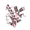
| Biological species |  | ||||||
| Method |  X-RAY DIFFRACTION / SYNCHROTRON / MOLECULAR REPLACEMENT / Resolution: 2 Å X-RAY DIFFRACTION / SYNCHROTRON / MOLECULAR REPLACEMENT / Resolution: 2 Å | ||||||
 Authors Authors | Ballut, L. / Aghajari, N. | ||||||
 Citation Citation | Journal: J. Mol. Biol. / Year: 2017 Title: Expanding the Kinome World: A New Protein Kinase Family Widely Conserved in Bacteria. Authors: Nguyen, H.A. / El Khoury, T. / Guiral, S. / Laaberki, M.H. / Candusso, M.P. / Galisson, F. / Foucher, A.E. / Kesraoui, S. / Ballut, L. / Vallet, S. / Orelle, C. / Zucchini, L. / Martin, J. / ...Authors: Nguyen, H.A. / El Khoury, T. / Guiral, S. / Laaberki, M.H. / Candusso, M.P. / Galisson, F. / Foucher, A.E. / Kesraoui, S. / Ballut, L. / Vallet, S. / Orelle, C. / Zucchini, L. / Martin, J. / Page, A. / Attieh, J. / Aghajari, N. / Grangeasse, C. / Jault, J.M. | ||||||
| History |
|
- Structure visualization
| Structure viewer | Molecule: MolmilJmol/JSmol |
|---|
- Downloads & links
-Download
| PDBx/mmCIF format | 5np9.cif.gz | 81.8 KB | Display | PDBx/mmCIF format |
|---|---|---|---|---|
| PDB format | pdb5np9.ent.gz | 60.3 KB | Display | PDB format |
| PDBx/mmJSON format | 5np9.json.gz | Tree view | PDBx/mmJSON format | |
| Others |  Other downloads Other downloads |
-Validation report
| Arichive directory | https://data.pdbj.org/pub/pdb/validation_reports/np/5np9ftp://data.pdbj.org/pub/pdb/validation_reports/np/5np9 | HTTPS FTP |
|---|
-Related structure data
| Related structure data | 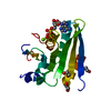 5mvrSC S: Starting model for refinement C: citing same article ( |
|---|---|
| Similar structure data |


-Links
 PDBj
PDBj- Assembly
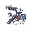
| Deposited unit | 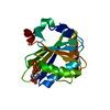
| ||||||||
|---|---|---|---|---|---|---|---|---|---|
| 1 |
| ||||||||
| Unit cell |
|
-Components
| #1: Protein | Mass: 17927.225 Da / Num. of mol.: 1 Source method: isolated from a genetically manipulated source Details: construction different fron that used for pdb entry 5MVR There is a flexible region from residue 87-89 and no electron density is visible for residues 87 and 88. As concnerns residue 89, ...Details: construction different fron that used for pdb entry 5MVR There is a flexible region from residue 87-89 and no electron density is visible for residues 87 and 88. As concnerns residue 89, there is no density for the side-chain. Hereafter, the sequence is complete until residue 154 (incl.) and from then on there is no more electron density. Source: (gene. exp.) |
|---|---|
| #2: Chemical | ChemComp-ADP /   Mass: 427.201 Da / Num. of mol.: 1 / Source method: isolated from a natural source / Formula: C10H15N5O10P2 / Comment: ADP, energy-carrying molecule*YM Mass: 427.201 Da / Num. of mol.: 1 / Source method: isolated from a natural source / Formula: C10H15N5O10P2 / Comment: ADP, energy-carrying molecule*YM |
| #3: Chemical | ChemComp-MG /   Mass: 24.305 Da / Num. of mol.: 1 / Source method: obtained synthetically / Formula: Mg Mass: 24.305 Da / Num. of mol.: 1 / Source method: obtained synthetically / Formula: Mg |
| #4: Water | ChemComp-HOH /  Mass: 18.015 Da / Num. of mol.: 86 / Source method: isolated from a natural source / Formula: H2O Mass: 18.015 Da / Num. of mol.: 86 / Source method: isolated from a natural source / Formula: H2O |
-Experimental details
-Experiment
| Experiment | Method: X-RAY DIFFRACTION / Number of used crystals: 1 |
|---|
- Sample preparation
| Crystal | Density Matthews: 2.34 Å3/Da / Density % sol: 47.36 % |
|---|---|
| Crystal grow | Temperature: 292 K / Method: vapor diffusion, sitting drop / Details: 30% PEG 4000, 200 mM MgCl2 and 100 mM Tris pH 8.5 |
-Data collection
| Diffraction | Mean temperature: 100 K |
|---|---|
| Diffraction source | Source: SYNCHROTRON / Site: ESRF  / Beamline: ID29 / Wavelength: 0.97625 Å / Beamline: ID29 / Wavelength: 0.97625 Å |
| Detector | Type: DECTRIS PILATUS 6M / Detector: PIXEL / Date: Feb 3, 2017 |
| Radiation | Monochromator: Liquid nitrogen cooled silicon monochromator / Protocol: SINGLE WAVELENGTH / Monochromatic (M) / Laue (L): M / Scattering type: x-ray |
| Radiation wavelength | Wavelength: 0.97625 Å / Relative weight: 1 |
| Reflection | Resolution: 2.05→34.06 Å / Num. obs: 10574 / % possible obs: 97.1 % / Redundancy: 3.7 % / Net I/σ(I): 8.2 |
- Processing
| Software |
| ||||||||||||||||||||||||||||||||||||||||
|---|---|---|---|---|---|---|---|---|---|---|---|---|---|---|---|---|---|---|---|---|---|---|---|---|---|---|---|---|---|---|---|---|---|---|---|---|---|---|---|---|---|
| Refinement | Method to determine structure: MOLECULAR REPLACEMENT Starting model: 5MVR Resolution: 2→34.059 Å / SU ML: 0.25 / Cross valid method: FREE R-VALUE / σ(F): 1.35 / Phase error: 25.8 / Stereochemistry target values: ML
| ||||||||||||||||||||||||||||||||||||||||
| Solvent computation | Shrinkage radii: 0.9 Å / VDW probe radii: 1.11 Å / Solvent model: FLAT BULK SOLVENT MODEL | ||||||||||||||||||||||||||||||||||||||||
| Refinement step | Cycle: LAST / Resolution: 2→34.059 Å
| ||||||||||||||||||||||||||||||||||||||||
| Refine LS restraints |
| ||||||||||||||||||||||||||||||||||||||||
| LS refinement shell |
| ||||||||||||||||||||||||||||||||||||||||
| Refinement TLS params. | Method: refined / Origin x: 25.3866 Å / Origin y: 16.5229 Å / Origin z: -3.4841 Å
| ||||||||||||||||||||||||||||||||||||||||
| Refinement TLS group | Selection details: all |
