 Movie
Movie Controller

[English] 日本語
 Yorodumi
- PDB-3qzu: Crystal structure of Bacillus subtilis Lipase A 7-fold mutant; th... -
+ Open data

- Basic information
| Entry | Database: PDB / ID: 3qzu | ||||||
|---|---|---|---|---|---|---|---|
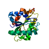
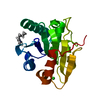
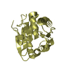
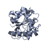
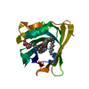
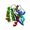
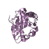
| Title | Crystal structure of Bacillus subtilis Lipase A 7-fold mutant; the outcome of directed evolution towards thermostability | ||||||
 Components Components | Lipase estA | ||||||
 Keywords Keywords | HYDROLASE / Alpha/beta hydrolase | ||||||
| Function / homology |  Function and homology information Function and homology informationlipase activity / triacylglycerol lipase / triacylglycerol lipase activity / lipid catabolic process / extracellular region Similarity search - Function | ||||||
| Biological species |  | ||||||
| Method |  X-RAY DIFFRACTION / SYNCHROTRON / MOLECULAR REPLACEMENT / Resolution: 1.85 Å X-RAY DIFFRACTION / SYNCHROTRON / MOLECULAR REPLACEMENT / Resolution: 1.85 Å | ||||||
 Authors Authors | Pijning, T. / Augustyniak, W. / Reetz, M.T. / Dijkstra, B.W. | ||||||
 Citation Citation | Journal: Protein Sci. / Year: 2012 Title: Biophysical characterization of mutants of Bacillus subtilis lipase evolved for thermostability: Factors contributing to increased activity retention. Authors: Augustyniak, W. / Brzezinska, A.A. / Pijning, T. / Wienk, H. / Boelens, R. / Dijkstra, B.W. / Reetz, M.T. | ||||||
| History |
|
- Structure visualization
| Structure viewer | Molecule: MolmilJmol/JSmol |
|---|
- Downloads & links
-Download
| PDBx/mmCIF format | 3qzu.cif.gz | 92.2 KB | Display | PDBx/mmCIF format |
|---|---|---|---|---|
| PDB format | pdb3qzu.ent.gz | 69.4 KB | Display | PDB format |
| PDBx/mmJSON format | 3qzu.json.gz | Tree view | PDBx/mmJSON format | |
| Others |  Other downloads Other downloads |
-Validation report
| Arichive directory | https://data.pdbj.org/pub/pdb/validation_reports/qz/3qzuftp://data.pdbj.org/pub/pdb/validation_reports/qz/3qzu | HTTPS FTP |
|---|
-Related structure data
| Related structure data | 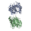 1i6wS S: Starting model for refinement |
|---|---|
| Similar structure data |


-Links
 PDBj
PDBj- Assembly
| Deposited unit | 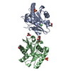
| ||||||||||||||||||
|---|---|---|---|---|---|---|---|---|---|---|---|---|---|---|---|---|---|---|---|
| 1 | 
| ||||||||||||||||||
| 2 | 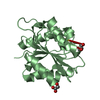
| ||||||||||||||||||
| Unit cell |
| ||||||||||||||||||
| Noncrystallographic symmetry (NCS) | NCS domain:
NCS domain segments: Component-ID: 1 / Ens-ID: 1 / Beg auth comp-ID: GLU / Beg label comp-ID: GLU / End auth comp-ID: ASN / End label comp-ID: ASN / Refine code: 5 / Auth seq-ID: 2 - 181 / Label seq-ID: 2 - 181
|
-Components
| #1: Protein | Mass: 19266.498 Da / Num. of mol.: 2 / Mutation: R33Q, D34N, K35D, K112D, M134D, Y139C, I157M Source method: isolated from a genetically manipulated source Source: (gene. exp.) Strain: 168 / Gene: BSU02700, estA, lip, lipA / Plasmid: pET22b / Production host: #2: Chemical |   Mass: 35.453 Da / Num. of mol.: 2 / Source method: obtained synthetically / Formula: Cl Mass: 35.453 Da / Num. of mol.: 2 / Source method: obtained synthetically / Formula: Cl#3: Chemical |   Mass: 96.063 Da / Num. of mol.: 2 / Source method: obtained synthetically / Formula: SO4 Mass: 96.063 Da / Num. of mol.: 2 / Source method: obtained synthetically / Formula: SO4#4: Chemical | ChemComp-GOL /   Mass: 92.094 Da / Num. of mol.: 5 / Source method: obtained synthetically / Formula: C3H8O3 Mass: 92.094 Da / Num. of mol.: 5 / Source method: obtained synthetically / Formula: C3H8O3#5: Water | ChemComp-HOH / |  Mass: 18.015 Da / Num. of mol.: 482 / Source method: isolated from a natural source / Formula: H2O Mass: 18.015 Da / Num. of mol.: 482 / Source method: isolated from a natural source / Formula: H2OHas protein modification | Y | |
|---|
-Experimental details
-Experiment
| Experiment | Method: X-RAY DIFFRACTION / Number of used crystals: 1 |
|---|
- Sample preparation
| Crystal | Density Matthews: 2.66 Å3/Da / Density % sol: 53.78 % |
|---|---|
| Crystal grow | Temperature: 293 K / Method: vapor diffusion, hanging drop / pH: 6 Details: 24% (w/v) PEG 3350, 0.2 M (NH4)2)SO4, 0.1 M BisTris-HCl, 0.03 M BES-NaOH, 0.03% (w/v) NaN3, pH 6.0, VAPOR DIFFUSION, HANGING DROP, temperature 293K |
-Data collection
| Diffraction | Mean temperature: 100 K |
|---|---|
| Diffraction source | Source: SYNCHROTRON / Site: ESRF  / Beamline: ID23-2 / Wavelength: 0.8726 Å / Beamline: ID23-2 / Wavelength: 0.8726 Å |
| Detector | Type: MARMOSAIC 225 mm CCD / Detector: CCD / Date: Jun 10, 2010 / Details: mirrors |
| Radiation | Protocol: SINGLE WAVELENGTH / Monochromatic (M) / Laue (L): M / Scattering type: x-ray |
| Radiation wavelength | Wavelength: 0.8726 Å / Relative weight: 1 |
| Reflection | Resolution: 1.71→45.51 Å / Num. all: 43880 / Num. obs: 43134 / % possible obs: 98.3 % / Observed criterion σ(I): 3 / Redundancy: 4.1 % / Biso Wilson estimate: 16.9 Å2 / Rmerge(I) obs: 0.159 / Net I/σ(I): 6.8 |
| Reflection shell | Resolution: 1.71→1.81 Å / Redundancy: 3.3 % / Rmerge(I) obs: 0.822 / Mean I/σ(I) obs: 1.5 / Num. unique all: 18453 / % possible all: 88.2 |
- Processing
| Software | Name: REFMAC / Version: 5.5.0102 / Classification: refinement | |||||||||||||||||||||||||||||||||||||||||||||||||||||||||||||||||
|---|---|---|---|---|---|---|---|---|---|---|---|---|---|---|---|---|---|---|---|---|---|---|---|---|---|---|---|---|---|---|---|---|---|---|---|---|---|---|---|---|---|---|---|---|---|---|---|---|---|---|---|---|---|---|---|---|---|---|---|---|---|---|---|---|---|---|
| Refinement | Method to determine structure: MOLECULAR REPLACEMENT Starting model: PDB ENTRY 1I6W Resolution: 1.85→76.16 Å / Cor.coef. Fo:Fc: 0.948 / Cor.coef. Fo:Fc free: 0.907 / SU B: 2.725 / SU ML: 0.083 / Isotropic thermal model: isotropic / Cross valid method: THROUGHOUT / ESU R Free: 0.134 / Stereochemistry target values: MAXIMUM LIKELIHOOD / Details: HYDROGENS HAVE BEEN ADDED IN THE RIDING POSITIONS
| |||||||||||||||||||||||||||||||||||||||||||||||||||||||||||||||||
| Solvent computation | Ion probe radii: 0.8 Å / Shrinkage radii: 0.8 Å / VDW probe radii: 1.4 Å / Solvent model: MASK | |||||||||||||||||||||||||||||||||||||||||||||||||||||||||||||||||
| Displacement parameters | Biso mean: 13.792 Å2
| |||||||||||||||||||||||||||||||||||||||||||||||||||||||||||||||||
| Refinement step | Cycle: LAST / Resolution: 1.85→76.16 Å
| |||||||||||||||||||||||||||||||||||||||||||||||||||||||||||||||||
| Refine LS restraints |
| |||||||||||||||||||||||||||||||||||||||||||||||||||||||||||||||||
| Refine LS restraints NCS | Dom-ID: 1 / Auth asym-ID: A / Ens-ID: 1 / Refine-ID: X-RAY DIFFRACTION
| |||||||||||||||||||||||||||||||||||||||||||||||||||||||||||||||||
| LS refinement shell | Resolution: 1.85→1.898 Å / Total num. of bins used: 20
|
