 Movie
Movie Controller

[English] 日本語
 Yorodumi
- PDB-2qxt: Crystal Structure Analysis of the Bacillus subtilis lipase crysta... -
+ Open data

- Basic information
| Entry | Database: PDB / ID: 2qxt | ||||||
|---|---|---|---|---|---|---|---|
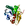
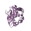
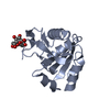
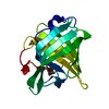
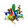
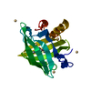
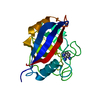
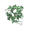
| Title | Crystal Structure Analysis of the Bacillus subtilis lipase crystallized at pH 4.5 | ||||||
 Components Components | Lipase | ||||||
 Keywords Keywords | HYDROLASE / alpha/beta hydrolase fold / Lipid degradation / Secreted | ||||||
| Function / homology |  Function and homology information Function and homology informationlipase activity / triacylglycerol lipase / triacylglycerol lipase activity / lipid catabolic process / extracellular region Similarity search - Function | ||||||
| Biological species |  | ||||||
| Method |  X-RAY DIFFRACTION / MOLECULAR REPLACEMENT / Resolution: 2 Å X-RAY DIFFRACTION / MOLECULAR REPLACEMENT / Resolution: 2 Å | ||||||
 Authors Authors | Rajakumara, E. / Sankaranarayanan, R. | ||||||
 Citation Citation | Journal: Biochim.Biophys.Acta / Year: 2008 Title: Structural basis for the remarkable stability of Bacillus subtilis lipase (Lip A) at low pH Authors: Rajakumara, E. / Acharya, P. / Ahmad, S. / Sankaranaryanan, R. / Rao, N.M. | ||||||
| History |
|
- Structure visualization
| Structure viewer | Molecule: MolmilJmol/JSmol |
|---|
- Downloads & links
-Download
| PDBx/mmCIF format | 2qxt.cif.gz | 83.1 KB | Display | PDBx/mmCIF format |
|---|---|---|---|---|
| PDB format | pdb2qxt.ent.gz | 63.5 KB | Display | PDB format |
| PDBx/mmJSON format | 2qxt.json.gz | Tree view | PDBx/mmJSON format | |
| Others |  Other downloads Other downloads |
-Validation report
| Arichive directory | https://data.pdbj.org/pub/pdb/validation_reports/qx/2qxtftp://data.pdbj.org/pub/pdb/validation_reports/qx/2qxt | HTTPS FTP |
|---|
-Related structure data
| Related structure data |  2qxuC 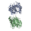 1i6wS C: citing same article ( S: Starting model for refinement |
|---|---|
| Similar structure data |


-Links
 PDBj
PDBj- Assembly
| Deposited unit | 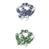
| ||||||||
|---|---|---|---|---|---|---|---|---|---|
| 1 | 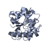
| ||||||||
| 2 | 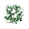
| ||||||||
| 3 | 
| ||||||||
| Unit cell |
|
-Components
| #1: Protein | Mass: 19182.643 Da / Num. of mol.: 2 Source method: isolated from a genetically manipulated source Source: (gene. exp.) #2: Water | ChemComp-HOH / |  Mass: 18.015 Da / Num. of mol.: 280 / Source method: isolated from a natural source / Formula: H2O Mass: 18.015 Da / Num. of mol.: 280 / Source method: isolated from a natural source / Formula: H2O |
|---|
-Experimental details
-Experiment
| Experiment | Method: X-RAY DIFFRACTION / Number of used crystals: 1 |
|---|
- Sample preparation
| Crystal | Density Matthews: 2.05 Å3/Da / Density % sol: 39.98 % |
|---|---|
| Crystal grow | Temperature: 298 K / Method: vapor diffusion / pH: 4.5 Details: 26% PEG 4000, 50mM Sodium Citrate pH 4.5, 50mM Ammonium Sulfate, VAPOR DIFFUSION, temperature 298K |
-Data collection
| Diffraction | Mean temperature: 100 K |
|---|---|
| Diffraction source | Source: ROTATING ANODE / Type: RIGAKU RUH3R / Wavelength: 1.5418 Å |
| Detector | Type: MAR scanner 345 mm plate / Detector: IMAGE PLATE / Date: Sep 10, 2003 / Details: mirrors |
| Radiation | Monochromator: Osmic Mirror System / Protocol: SINGLE WAVELENGTH / Monochromatic (M) / Laue (L): M / Scattering type: x-ray |
| Radiation wavelength | Wavelength: 1.5418 Å / Relative weight: 1 |
| Reflection | Resolution: 2→25 Å / Num. all: 22197 / Num. obs: 21687 / % possible obs: 97.7 % / Observed criterion σ(F): 0 / Observed criterion σ(I): 0 / Redundancy: 4.2 % / Biso Wilson estimate: 17.3 Å2 / Rsym value: 0.106 / Net I/σ(I): 11.1 |
| Reflection shell | Resolution: 2→2.07 Å / Redundancy: 4 % / Mean I/σ(I) obs: 2.3 / Num. unique all: 2219 / Rsym value: 0.457 / % possible all: 95.4 |
- Processing
| Software |
| ||||||||||||||||||||||||||||||||||||
|---|---|---|---|---|---|---|---|---|---|---|---|---|---|---|---|---|---|---|---|---|---|---|---|---|---|---|---|---|---|---|---|---|---|---|---|---|---|
| Refinement | Method to determine structure: MOLECULAR REPLACEMENT Starting model: PDB entry 1I6W Resolution: 2→24.94 Å / Rfactor Rfree error: 0.008 / Data cutoff high absF: 2543611.47 / Data cutoff low absF: 0 / Isotropic thermal model: RESTRAINED / Cross valid method: THROUGHOUT / σ(F): 0 / σ(I): 0 / Stereochemistry target values: Engh & Huber
| ||||||||||||||||||||||||||||||||||||
| Solvent computation | Solvent model: FLAT MODEL / Bsol: 43.1144 Å2 / ksol: 0.347426 e/Å3 | ||||||||||||||||||||||||||||||||||||
| Displacement parameters | Biso mean: 20.6 Å2
| ||||||||||||||||||||||||||||||||||||
| Refine analyze |
| ||||||||||||||||||||||||||||||||||||
| Refinement step | Cycle: LAST / Resolution: 2→24.94 Å
| ||||||||||||||||||||||||||||||||||||
| Refine LS restraints |
| ||||||||||||||||||||||||||||||||||||
| LS refinement shell | Resolution: 2→2.07 Å / Rfactor Rfree error: 0.036 / Total num. of bins used: 10
| ||||||||||||||||||||||||||||||||||||
| Xplor file |
|
