 Movie
Movie Controller


+ Open data

- Basic information
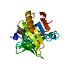
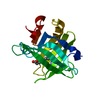
| Entry | Database: PDB / ID: 4dq3 | |||||||||
|---|---|---|---|---|---|---|---|---|---|---|
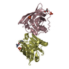
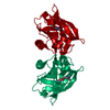
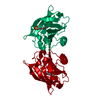
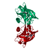
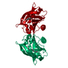
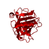
| Title | Bovine beta-lactoglobulin complex with oleic acid | |||||||||
 Components Components | Beta-lactoglobulin | |||||||||
 Keywords Keywords | TRANSPORT PROTEIN / lipocalin / transoprt protein / bovine milk | |||||||||
| Function / homology |  Function and homology information Function and homology informationretinol binding / long-chain fatty acid binding / extracellular region / identical protein binding Similarity search - Function | |||||||||
| Biological species |  | |||||||||
| Method |  X-RAY DIFFRACTION / MOLECULAR REPLACEMENT / Resolution: 2.1 Å X-RAY DIFFRACTION / MOLECULAR REPLACEMENT / Resolution: 2.1 Å | |||||||||
 Authors Authors | Loch, J.I. / Lewinski, K. | |||||||||
 Citation Citation | Journal: Int.J.Biol.Macromol. / Year: 2013 Title: Binding of 18-carbon unsaturated fatty acids to bovine beta-lactoglobulin--structural and thermodynamic studies. Authors: Loch, J.I. / Bonarek, P. / Polit, A. / Ries, D. / Dziedzicka-Wasylewska, M. / Lewinski, K. | |||||||||
| History |
|
- Structure visualization
| Structure viewer | Molecule: MolmilJmol/JSmol |
|---|
- Downloads & links
-Download
| PDBx/mmCIF format | 4dq3.cif.gz | 47.9 KB | Display | PDBx/mmCIF format |
|---|---|---|---|---|
| PDB format | pdb4dq3.ent.gz | 33.1 KB | Display | PDB format |
| PDBx/mmJSON format | 4dq3.json.gz | Tree view | PDBx/mmJSON format | |
| Others |  Other downloads Other downloads |
-Validation report
| Arichive directory | https://data.pdbj.org/pub/pdb/validation_reports/dq/4dq3ftp://data.pdbj.org/pub/pdb/validation_reports/dq/4dq3 | HTTPS FTP |
|---|
-Related structure data
| Related structure data | 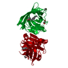 4dq4C 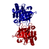 1bsyS S: Starting model for refinement C: citing same article ( |
|---|---|
| Similar structure data |


-Links
 PDBj
PDBj


- Assembly
| Deposited unit | 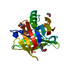
| ||||||||
|---|---|---|---|---|---|---|---|---|---|
| 1 | 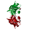
| ||||||||
| 2 |
| ||||||||
| Unit cell |
|
-Components
| #1: Protein | Mass: 18301.174 Da / Num. of mol.: 1 / Fragment: UNP residues 17-178 / Source method: isolated from a natural source / Source: (natural) |
|---|---|
| #2: Chemical | ChemComp-OLA /   Mass: 282.461 Da / Num. of mol.: 1 / Source method: obtained synthetically / Formula: C18H34O2 Mass: 282.461 Da / Num. of mol.: 1 / Source method: obtained synthetically / Formula: C18H34O2 |
| #3: Chemical | ChemComp-EOH /   Mass: 46.068 Da / Num. of mol.: 1 / Source method: obtained synthetically / Formula: C2H6O Mass: 46.068 Da / Num. of mol.: 1 / Source method: obtained synthetically / Formula: C2H6O |
| #4: Chemical | ChemComp-GOL /   Mass: 92.094 Da / Num. of mol.: 1 / Source method: obtained synthetically / Formula: C3H8O3 Mass: 92.094 Da / Num. of mol.: 1 / Source method: obtained synthetically / Formula: C3H8O3 |
| #5: Water | ChemComp-HOH /  Mass: 18.015 Da / Num. of mol.: 68 / Source method: isolated from a natural source / Formula: H2O Mass: 18.015 Da / Num. of mol.: 68 / Source method: isolated from a natural source / Formula: H2O |
| Has protein modification | Y |
-Experimental details
-Experiment
| Experiment | Method: X-RAY DIFFRACTION / Number of used crystals: 1 |
|---|
- Sample preparation
| Crystal | Density Matthews: 2.51 Å3/Da / Density % sol: 51.09 % |
|---|---|
| Crystal grow | Temperature: 293 K / Method: vapor diffusion, hanging drop / pH: 7.5 Details: 0.1M Tris-HCl buffer, 1.34M tri-sodium citrate, 1mM oleic acid in ethanol, pH 7.5, VAPOR DIFFUSION, HANGING DROP, temperature 293K |
-Data collection
| Diffraction | Mean temperature: 110 K |
|---|---|
| Diffraction source | Source: SEALED TUBE / Type: OXFORD DIFFRACTION ENHANCE ULTRA / Wavelength: 1.54 Å |
| Detector | Type: AGILENT ATLAS CCD / Detector: CCD / Date: Dec 10, 2009 |
| Radiation | Protocol: SINGLE WAVELENGTH / Monochromatic (M) / Laue (L): M / Scattering type: x-ray |
| Radiation wavelength | Wavelength: 1.54 Å / Relative weight: 1 |
| Reflection | Resolution: 2.1→14.87 Å / Num. all: 11278 / Num. obs: 11154 / % possible obs: 98.9 % / Observed criterion σ(F): 2 / Observed criterion σ(I): 2 / Rmerge(I) obs: 0.038 / Net I/σ(I): 13.3 |
| Reflection shell | Resolution: 2.1→2.21 Å / Rmerge(I) obs: 0.155 / Mean I/σ(I) obs: 3.5 / % possible all: 96.9 |
- Processing
| Software |
| ||||||||||||||||||||||||||||||||||||||||||||||||||||||||||||||||||||||||||||||||||||||||||
|---|---|---|---|---|---|---|---|---|---|---|---|---|---|---|---|---|---|---|---|---|---|---|---|---|---|---|---|---|---|---|---|---|---|---|---|---|---|---|---|---|---|---|---|---|---|---|---|---|---|---|---|---|---|---|---|---|---|---|---|---|---|---|---|---|---|---|---|---|---|---|---|---|---|---|---|---|---|---|---|---|---|---|---|---|---|---|---|---|---|---|---|
| Refinement | Method to determine structure: MOLECULAR REPLACEMENT Starting model: PDB ENTRY 1BSY Resolution: 2.1→14.87 Å / Cor.coef. Fo:Fc: 0.933 / Cor.coef. Fo:Fc free: 0.903 / SU B: 6.855 / SU ML: 0.183 / Cross valid method: THROUGHOUT / ESU R: 0.28 / ESU R Free: 0.234 / Stereochemistry target values: MAXIMUM LIKELIHOOD / Details: HYDROGENS HAVE BEEN ADDED IN THE RIDING POSITIONS
| ||||||||||||||||||||||||||||||||||||||||||||||||||||||||||||||||||||||||||||||||||||||||||
| Solvent computation | Ion probe radii: 0.8 Å / Shrinkage radii: 0.8 Å / VDW probe radii: 1.2 Å / Solvent model: MASK | ||||||||||||||||||||||||||||||||||||||||||||||||||||||||||||||||||||||||||||||||||||||||||
| Displacement parameters | Biso mean: 29.911 Å2
| ||||||||||||||||||||||||||||||||||||||||||||||||||||||||||||||||||||||||||||||||||||||||||
| Refinement step | Cycle: LAST / Resolution: 2.1→14.87 Å
| ||||||||||||||||||||||||||||||||||||||||||||||||||||||||||||||||||||||||||||||||||||||||||
| Refine LS restraints |
| ||||||||||||||||||||||||||||||||||||||||||||||||||||||||||||||||||||||||||||||||||||||||||
| LS refinement shell | Resolution: 2.1→2.154 Å / Total num. of bins used: 20
|
