 Movie
Movie Controller

[English] 日本語
 Yorodumi
- PDB-1dyw: Biochemical and structural characterization of a divergent loop c... -
+ Open data

- Basic information
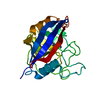
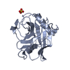
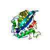
| Entry | Database: PDB / ID: 1dyw | |||||||||
|---|---|---|---|---|---|---|---|---|---|---|
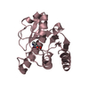
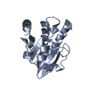
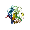
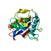
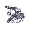
| Title | Biochemical and structural characterization of a divergent loop cyclophilin from Caenorhabditis elegans | |||||||||
 Components Components | CYCLOPHILIN 3 | |||||||||
 Keywords Keywords | ISOMERASE(PEPTIDYL-PROLYL CIS-TRANS) / ISOMERASE / ROTAMASE | |||||||||
| Function / homology |  Function and homology information Function and homology informationcyclosporin A binding / peptidylprolyl isomerase / peptidyl-prolyl cis-trans isomerase activity / protein folding / cytoplasm Similarity search - Function | |||||||||
| Biological species |  | |||||||||
| Method |  X-RAY DIFFRACTION / MOLECULAR REPLACEMENT / Resolution: 1.8 Å X-RAY DIFFRACTION / MOLECULAR REPLACEMENT / Resolution: 1.8 Å | |||||||||
 Authors Authors | Dornan, J. / Page, A.P. / Taylor, P. / Wu, S.Y. / Winter, A.D. / Husi, H. / Walkinshaw, M.D. | |||||||||
 Citation Citation | Journal: J.Biol.Chem. / Year: 1999 Title: Biochemical and Structural Characterization of a Divergent Loop Cyclophilin from Caenorhabditis Elegans Authors: Dornan, J. / Page, A.P. / Taylor, P. / Wu, S.Y. / Winter, A.D. / Husi, H. / Walkinshaw, M.D. #1: Journal: J.Mol.Biol. / Year: 1993Title: X-Ray Structure of a Monomeric Cyclophilin Authors: Mikol, V. / Kallen, J. / Pfluegl, G. / Walkinshaw, M.D. | |||||||||
| History |
|
- Structure visualization
| Structure viewer | Molecule: MolmilJmol/JSmol |
|---|
- Downloads & links
-Download
| PDBx/mmCIF format | 1dyw.cif.gz | 50.9 KB | Display | PDBx/mmCIF format |
|---|---|---|---|---|
| PDB format | pdb1dyw.ent.gz | 35.2 KB | Display | PDB format |
| PDBx/mmJSON format | 1dyw.json.gz | Tree view | PDBx/mmJSON format | |
| Others |  Other downloads Other downloads |
-Validation report
| Arichive directory | https://data.pdbj.org/pub/pdb/validation_reports/dy/1dywftp://data.pdbj.org/pub/pdb/validation_reports/dy/1dyw | HTTPS FTP |
|---|
-Related structure data
| Related structure data | 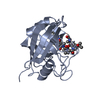 1cwaS S: Starting model for refinement |
|---|---|
| Similar structure data |


-Links
 PDBj
PDBj

- Assembly
| Deposited unit | 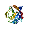
| ||||||||
|---|---|---|---|---|---|---|---|---|---|
| 1 |
| ||||||||
| Unit cell |
| ||||||||
| Details | BIOLOGICAL_UNIT: MONOMER |
-Components
| #1: Protein | Mass: 18576.182 Da / Num. of mol.: 1 Source method: isolated from a genetically manipulated source Source: (gene. exp.)  |
|---|---|
| #2: Water | ChemComp-HOH /  Mass: 18.015 Da / Num. of mol.: 252 / Source method: isolated from a natural source / Formula: H2O Mass: 18.015 Da / Num. of mol.: 252 / Source method: isolated from a natural source / Formula: H2O |
-Experimental details
-Experiment
| Experiment | Method: X-RAY DIFFRACTION / Number of used crystals: 1 |
|---|
- Sample preparation
| Crystal | Density Matthews: 3.05 Å3/Da / Density % sol: 59.32 % | ||||||||||||||||||||||||||||||
|---|---|---|---|---|---|---|---|---|---|---|---|---|---|---|---|---|---|---|---|---|---|---|---|---|---|---|---|---|---|---|---|
| Crystal grow | pH: 5.6 / Details: MPEG5000, SODIUM CITRATE, PH 5.6 | ||||||||||||||||||||||||||||||
| Crystal grow | *PLUS Temperature: 18 ℃ / Method: vapor diffusion, hanging drop | ||||||||||||||||||||||||||||||
| Components of the solutions | *PLUS
|
-Data collection
| Diffraction | Mean temperature: 100 K |
|---|---|
| Diffraction source | Source: ROTATING ANODE / Type: ENRAF-NONIUS FR571 / Wavelength: 1.5418 |
| Detector | Type: MARRESEARCH / Detector: IMAGE PLATE / Details: COLLIMATOR |
| Radiation | Monochromator: GRAPHITE(002) / Protocol: SINGLE WAVELENGTH / Monochromatic (M) / Laue (L): M / Scattering type: x-ray |
| Radiation wavelength | Wavelength: 1.5418 Å / Relative weight: 1 |
| Reflection | Resolution: 1.8→15 Å / Num. obs: 21337 / % possible obs: 97 % / Redundancy: 6.59 % / Rsym value: 0.083 / Net I/σ(I): 11.75 |
| Reflection shell | Resolution: 1.8→1.83 Å / Mean I/σ(I) obs: 2.587 / Rsym value: 0.278 / % possible all: 96.2 |
| Reflection | *PLUS Rmerge(I) obs: 0.083 |
| Reflection shell | *PLUS % possible obs: 96.2 % / Rmerge(I) obs: 0.278 |
- Processing
| Software |
| |||||||||||||||||||||||||||||||||
|---|---|---|---|---|---|---|---|---|---|---|---|---|---|---|---|---|---|---|---|---|---|---|---|---|---|---|---|---|---|---|---|---|---|---|
| Refinement | Method to determine structure: MOLECULAR REPLACEMENT Starting model: 1CWA Resolution: 1.8→15 Å / Num. parameters: 6247 / Num. restraintsaints: 5295 / Cross valid method: FREE R-VALUE / σ(F): 0 / Stereochemistry target values: ENGH AND HUBER Details: THE C-TERMINAL RESIDUE WAS NOT SEEN IN THE DENSITY MAPS
| |||||||||||||||||||||||||||||||||
| Solvent computation | Solvent model: MOEWS & KRETSINGER, J.MOL.BIOL.91(1973)201-2 | |||||||||||||||||||||||||||||||||
| Refine analyze | Num. disordered residues: 0 / Occupancy sum hydrogen: 0 / Occupancy sum non hydrogen: 1561 | |||||||||||||||||||||||||||||||||
| Refinement step | Cycle: LAST / Resolution: 1.8→15 Å
| |||||||||||||||||||||||||||||||||
| Refine LS restraints |
| |||||||||||||||||||||||||||||||||
| Software | *PLUS Name: SHELXL-97 / Classification: refinement | |||||||||||||||||||||||||||||||||
| Refinement | *PLUS | |||||||||||||||||||||||||||||||||
| Solvent computation | *PLUS | |||||||||||||||||||||||||||||||||
| Displacement parameters | *PLUS Biso mean: 30.37 Å2 |
