 Movie
Movie Controller

[English] 日本語
 Yorodumi
- PDB-5crx: ASYMMETRIC DNA-BENDING IN THE CRE-LOXP SITE-SPECIFIC RECOMBINATIO... -
+ Open data

- Basic information
| Entry | Database: PDB / ID: 5crx | ||||||
|---|---|---|---|---|---|---|---|
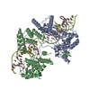
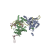
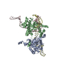
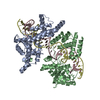
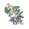
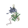
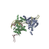
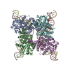
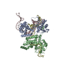
| Title | ASYMMETRIC DNA-BENDING IN THE CRE-LOXP SITE-SPECIFIC RECOMBINATION SYNAPSE | ||||||
 Components Components |
| ||||||
 Keywords Keywords | PROTEIN/DNA / CRE RECOMBINASE / DNA BENDING / SITE SPECIFIC RECOMBINATION / PROTEIN-DNA INTERACTION / PROTEIN-DNA complex | ||||||
| Function / homology |  Function and homology information Function and homology information | ||||||
| Biological species |  Enterobacteria phage P1 (virus) Enterobacteria phage P1 (virus) | ||||||
| Method |  X-RAY DIFFRACTION / SYNCHROTRON / MOLECULAR REPLACEMENT / Resolution: 2.7 Å X-RAY DIFFRACTION / SYNCHROTRON / MOLECULAR REPLACEMENT / Resolution: 2.7 Å | ||||||
 Authors Authors | Guo, F. / Gopaul, D.N. / Van Duyne, G.D. | ||||||
 Citation Citation | Journal: Proc.Natl.Acad.Sci.USA / Year: 1999 Title: Asymmetric DNA bending in the Cre-loxP site-specific recombination synapse. Authors: Guo, F. / Gopaul, D.N. / Van Duyne, G.D. #1: Journal: Embo J. / Year: 1998Title: Structure of the Holliday junction intermediate in Cre-loxP site-specific recombination. Authors: Gopaul, D.N. / Guo, F. / Van Duyne, G.D. #2: Journal: Nature / Year: 1997Title: Structure of Cre recombinase complexed with DNA in a site-specific recombination synapse. Authors: Guo, F. / Gopaul, D.N. / van Duyne, G.D. | ||||||
| History |
|
- Structure visualization
| Structure viewer | Molecule: MolmilJmol/JSmol |
|---|
- Downloads & links
-Download
| PDBx/mmCIF format | 5crx.cif.gz | 174.3 KB | Display | PDBx/mmCIF format |
|---|---|---|---|---|
| PDB format | pdb5crx.ent.gz | 133.6 KB | Display | PDB format |
| PDBx/mmJSON format | 5crx.json.gz | Tree view | PDBx/mmJSON format | |
| Others |  Other downloads Other downloads |
-Validation report
| Arichive directory | https://data.pdbj.org/pub/pdb/validation_reports/cr/5crxftp://data.pdbj.org/pub/pdb/validation_reports/cr/5crx | HTTPS FTP |
|---|
-Related structure data
| Related structure data |  4crxC 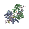 1crxS S: Starting model for refinement C: citing same article ( |
|---|---|
| Similar structure data |

-Links
 PDBj
PDBj


- Assembly
| Deposited unit | 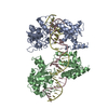
| ||||||||
|---|---|---|---|---|---|---|---|---|---|
| 1 | 
| ||||||||
| Unit cell |
|
-Components
| #1: DNA chain | Mass: 10759.968 Da / Num. of mol.: 2 / Source method: obtained synthetically #2: Protein | Mass: 38579.164 Da / Num. of mol.: 2 / Mutation: Y324F Source method: isolated from a genetically manipulated source Source: (gene. exp.) Enterobacteria phage P1 (virus) / Genus: P1-like viruses / Gene: CRE / Plasmid: PET21A / Species (production host): Escherichia coli / Cellular location (production host): CYTOPLASMIC / Production host:  #3: Water | ChemComp-HOH / |  Mass: 18.015 Da / Num. of mol.: 314 / Source method: isolated from a natural source / Formula: H2O Mass: 18.015 Da / Num. of mol.: 314 / Source method: isolated from a natural source / Formula: H2O |
|---|
-Experimental details
-Experiment
| Experiment | Method: X-RAY DIFFRACTION / Number of used crystals: 1 |
|---|
- Sample preparation
| Crystal | Density Matthews: 2.97 Å3/Da / Density % sol: 50 % | ||||||||||||||||||||||||||||||||||||||||||||||||||||||||||||||||||
|---|---|---|---|---|---|---|---|---|---|---|---|---|---|---|---|---|---|---|---|---|---|---|---|---|---|---|---|---|---|---|---|---|---|---|---|---|---|---|---|---|---|---|---|---|---|---|---|---|---|---|---|---|---|---|---|---|---|---|---|---|---|---|---|---|---|---|---|
| Crystal grow | pH: 5 / Details: pH 5.0 | ||||||||||||||||||||||||||||||||||||||||||||||||||||||||||||||||||
| Crystal | *PLUS | ||||||||||||||||||||||||||||||||||||||||||||||||||||||||||||||||||
| Crystal grow | *PLUS Temperature: 18 ℃ / pH: 5.6 / Method: vapor diffusion, hanging drop | ||||||||||||||||||||||||||||||||||||||||||||||||||||||||||||||||||
| Components of the solutions | *PLUS
|
-Data collection
| Diffraction | Mean temperature: 100 K |
|---|---|
| Diffraction source | Source: SYNCHROTRON / Site: CHESS  / Beamline: F2 / Wavelength: 0.9188 / Beamline: F2 / Wavelength: 0.9188 |
| Detector | Type: ADSC / Detector: CCD / Date: Dec 1, 1997 / Details: MIRRORS |
| Radiation | Monochromator: SI / Protocol: SINGLE WAVELENGTH / Monochromatic (M) / Laue (L): M / Scattering type: x-ray |
| Radiation wavelength | Wavelength: 0.9188 Å / Relative weight: 1 |
| Reflection | Resolution: 2.7→99 Å / Num. obs: 32242 / % possible obs: 97.3 % / Observed criterion σ(I): -3 / Redundancy: 4.8 % / Biso Wilson estimate: 63.4 Å2 / Rsym value: 0.069 / Net I/σ(I): 12.59 |
| Reflection shell | Resolution: 2.7→2.78 Å / Redundancy: 3.71 % / Mean I/σ(I) obs: 23.76 / Rsym value: 0.23 / % possible all: 93.9 |
| Reflection | *PLUS Rmerge(I) obs: 0.066 |
| Reflection shell | *PLUS % possible obs: 94 % / Rmerge(I) obs: 0.23 |
- Processing
| Software |
| ||||||||||||||||||||||||||||||||||||||||||||||||||||||||||||||||||||||||||||||||
|---|---|---|---|---|---|---|---|---|---|---|---|---|---|---|---|---|---|---|---|---|---|---|---|---|---|---|---|---|---|---|---|---|---|---|---|---|---|---|---|---|---|---|---|---|---|---|---|---|---|---|---|---|---|---|---|---|---|---|---|---|---|---|---|---|---|---|---|---|---|---|---|---|---|---|---|---|---|---|---|---|---|
| Refinement | Method to determine structure: MOLECULAR REPLACEMENT Starting model: 1CRX Resolution: 2.7→48 Å / Rfactor Rfree error: 0.017 / Data cutoff high absF: 1000000 / Data cutoff low absF: 0.001 / Isotropic thermal model: RESTRAINED / Cross valid method: THROUGHOUT / σ(F): 2
| ||||||||||||||||||||||||||||||||||||||||||||||||||||||||||||||||||||||||||||||||
| Displacement parameters | Biso mean: 50.4 Å2
| ||||||||||||||||||||||||||||||||||||||||||||||||||||||||||||||||||||||||||||||||
| Refine analyze |
| ||||||||||||||||||||||||||||||||||||||||||||||||||||||||||||||||||||||||||||||||
| Refinement step | Cycle: LAST / Resolution: 2.7→48 Å
| ||||||||||||||||||||||||||||||||||||||||||||||||||||||||||||||||||||||||||||||||
| Refine LS restraints |
| ||||||||||||||||||||||||||||||||||||||||||||||||||||||||||||||||||||||||||||||||
| LS refinement shell | Resolution: 2.7→2.82 Å / Rfactor Rfree error: 0.02 / Total num. of bins used: 8
| ||||||||||||||||||||||||||||||||||||||||||||||||||||||||||||||||||||||||||||||||
| Xplor file |
| ||||||||||||||||||||||||||||||||||||||||||||||||||||||||||||||||||||||||||||||||
| Software | *PLUS Name: X-PLOR / Version: 3.851 / Classification: refinement | ||||||||||||||||||||||||||||||||||||||||||||||||||||||||||||||||||||||||||||||||
| Refinement | *PLUS Highest resolution: 2.7 Å / Lowest resolution: 48 Å / σ(F): 2 / % reflection Rfree: 5 % | ||||||||||||||||||||||||||||||||||||||||||||||||||||||||||||||||||||||||||||||||
| Solvent computation | *PLUS | ||||||||||||||||||||||||||||||||||||||||||||||||||||||||||||||||||||||||||||||||
| Displacement parameters | *PLUS Biso mean: 50.4 Å2 | ||||||||||||||||||||||||||||||||||||||||||||||||||||||||||||||||||||||||||||||||
| Refine LS restraints | *PLUS
| ||||||||||||||||||||||||||||||||||||||||||||||||||||||||||||||||||||||||||||||||
| LS refinement shell | *PLUS Highest resolution: 2.7 Å / Rfactor Rfree: 0.437 / % reflection Rfree: 5 % / Rfactor Rwork: 0.337 |
