 Movie
Movie Controller

[English] 日本語
 Yorodumi
- PDB-1ma7: Crystal structure of Cre site-specific recombinase complexed with... -
+ Open data

- Basic information
| Entry | Database: PDB / ID: 1ma7 | ||||||
|---|---|---|---|---|---|---|---|
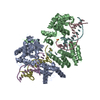
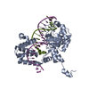
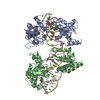
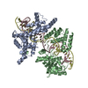
| Title | Crystal structure of Cre site-specific recombinase complexed with a mutant DNA substrate, LoxP-A8/T27 | ||||||
 Components Components |
| ||||||
 Keywords Keywords | HYDROLASE / LIGASE/DNA / Cre-Lox site-specific recombination / Specificity of protein-DNA interactions / protein-DNA complex / Tyrosine recombinase / LIGASE-DNA COMPLEX | ||||||
| Function / homology |  Function and homology information Function and homology information | ||||||
| Biological species |  Enterobacteria phage P1 (virus) Enterobacteria phage P1 (virus) | ||||||
| Method |  X-RAY DIFFRACTION / SYNCHROTRON / FOURIER SYNTHESIS / Resolution: 2.3 Å X-RAY DIFFRACTION / SYNCHROTRON / FOURIER SYNTHESIS / Resolution: 2.3 Å | ||||||
 Authors Authors | Martin, S.S. / Chu, V.C. / Baldwin, E.P. | ||||||
 Citation Citation | Journal: Biochemistry / Year: 2003 Title: Modulation of the active complex assembly and turnover rate by protein-DNA interactions in Cre-LoxP recombination Authors: Martin, S.S. / Chu, V.C. / Baldwin, E.P. #1: Journal: J.Mol.Biol. / Year: 2002Title: The order of strand exchanges in Cre-LoxP recombination and its basis suggested by the crystal structure of a Cre-LoxP Holliday junction complex Authors: Martin, S.S. / Pulido, E. / Chu, V.C. / Lechner, T.S. / Baldwin, E.P. #2: Journal: Nature / Year: 1997Title: Structure of Cre recombinase complexed with DNA in a site-specific recombination synapse Authors: Guo, F. / Gopaul, D. / van Duyne, G.D. #3: Journal: Annu.Rev.Biophys.Biomol.Struct. / Year: 2001Title: A structural view of cre-loxp site-specific recombination Authors: van Duyne, G.D. | ||||||
| History |
|
- Structure visualization
| Structure viewer | Molecule: MolmilJmol/JSmol |
|---|
- Downloads & links
-Download
| PDBx/mmCIF format | 1ma7.cif.gz | 181.9 KB | Display | PDBx/mmCIF format |
|---|---|---|---|---|
| PDB format | pdb1ma7.ent.gz | 139.9 KB | Display | PDB format |
| PDBx/mmJSON format | 1ma7.json.gz | Tree view | PDBx/mmJSON format | |
| Others |  Other downloads Other downloads |
-Validation report
| Arichive directory | https://data.pdbj.org/pub/pdb/validation_reports/ma/1ma7ftp://data.pdbj.org/pub/pdb/validation_reports/ma/1ma7 | HTTPS FTP |
|---|
-Related structure data
| Related structure data |  1kbuS S: Starting model for refinement |
|---|---|
| Similar structure data |


-Links
 PDBj
PDBj


- Assembly
| Deposited unit | 
| ||||||||
|---|---|---|---|---|---|---|---|---|---|
| 1 | 
| ||||||||
| Unit cell |
| ||||||||
| Details | THE SECOND HALF OF THE TETRAMERIC BIOLOGICAL ASSEMBLY IS GENERATED BY THE CRYSTALLOGRAPHIC TWO-FOLD AXIS: X, -Y, -Z, AND A TRANSLATION OF 0, 1, 1. |
-Components
| #1: DNA chain | Mass: 10469.798 Da / Num. of mol.: 1 / Fragment: UPPER STRAND / Mutation: C8A,G27T / Source method: obtained synthetically | ||
|---|---|---|---|
| #2: DNA chain | Mass: 10438.788 Da / Num. of mol.: 1 / Fragment: LOWER STRAND / Mutation: C8A,G27T / Source method: obtained synthetically | ||
| #3: Protein | Mass: 39424.047 Da / Num. of mol.: 2 Source method: isolated from a genetically manipulated source Source: (gene. exp.) Enterobacteria phage P1 (virus) / Genus: P1-like viruses / Plasmid: pET28b(+) / Species (production host): Escherichia coli / Production host:  #4: Water | ChemComp-HOH / |  Mass: 18.015 Da / Num. of mol.: 283 / Source method: isolated from a natural source / Formula: H2O Mass: 18.015 Da / Num. of mol.: 283 / Source method: isolated from a natural source / Formula: H2O |
-Experimental details
-Experiment
| Experiment | Method: X-RAY DIFFRACTION / Number of used crystals: 1 |
|---|
- Sample preparation
| Crystal | Density Matthews: 2.95 Å3/Da / Density % sol: 58.24 % | |||||||||||||||||||||||||||||||||||
|---|---|---|---|---|---|---|---|---|---|---|---|---|---|---|---|---|---|---|---|---|---|---|---|---|---|---|---|---|---|---|---|---|---|---|---|---|
| Crystal grow | Temperature: 294 K / Method: vapor diffusion, hanging drop / pH: 5.25 Details: 2,4-methylpentanediol, sodium acetate, calcium chloride, pH 5.25, VAPOR DIFFUSION, HANGING DROP, temperature 294K | |||||||||||||||||||||||||||||||||||
| Components of the solutions |
| |||||||||||||||||||||||||||||||||||
| Crystal grow | *PLUS Temperature: 21 ℃ / Method: vapor diffusion, hanging drop | |||||||||||||||||||||||||||||||||||
| Components of the solutions | *PLUS
|
-Data collection
| Diffraction | Mean temperature: 100 K |
|---|---|
| Diffraction source | Source: SYNCHROTRON / Site: SSRL  / Beamline: BL9-2 / Wavelength: 0.98 Å / Beamline: BL9-2 / Wavelength: 0.98 Å |
| Detector | Type: ADSC QUANTUM 4 / Detector: CCD / Date: Jun 28, 2000 |
| Radiation | Protocol: SINGLE WAVELENGTH / Monochromatic (M) / Laue (L): M / Scattering type: x-ray |
| Radiation wavelength | Wavelength: 0.98 Å / Relative weight: 1 |
| Reflection | Resolution: 2.3→30 Å / Num. all: 54160 / Num. obs: 51997 / % possible obs: 97 % / Observed criterion σ(F): -3 / Observed criterion σ(I): -3 / Redundancy: 3.3 % / Biso Wilson estimate: 47 Å2 / Rmerge(I) obs: 0.034 / Net I/σ(I): 21 |
| Reflection shell | Resolution: 2.3→2.35 Å / Redundancy: 2.9 % / Rmerge(I) obs: 0.275 / Mean I/σ(I) obs: 3.8 / Num. unique all: 3473 / % possible all: 97.7 |
| Reflection | *PLUS % possible obs: 96.5 % / Num. measured all: 168540 |
| Reflection shell | *PLUS % possible obs: 97.7 % / Num. unique obs: 3473 |
- Processing
| Software |
| |||||||||||||||||||||||||
|---|---|---|---|---|---|---|---|---|---|---|---|---|---|---|---|---|---|---|---|---|---|---|---|---|---|---|
| Refinement | Method to determine structure: FOURIER SYNTHESIS Starting model: PDB entry 1KBU with mutated basepairs, side chains Arg259, Asp262, and Asp266 omitted Resolution: 2.3→5 Å Isotropic thermal model: No B value correction in scaling Fcalc to Fobs during refinement. Final R calculation with K=1.2137 B=0.44240 (TNT) σ(F): 0 / Stereochemistry target values: Engh & Huber
| |||||||||||||||||||||||||
| Displacement parameters | Biso mean: 57 Å2 | |||||||||||||||||||||||||
| Refine analyze | Luzzati d res low obs: 5 Å / Luzzati sigma a obs: 0.56 Å | |||||||||||||||||||||||||
| Refinement step | Cycle: LAST / Resolution: 2.3→5 Å
| |||||||||||||||||||||||||
| Refine LS restraints |
| |||||||||||||||||||||||||
| LS refinement shell | Resolution: 2.3→2.38 Å /
| |||||||||||||||||||||||||
| Refinement | *PLUS Num. reflection obs: 43981 / Num. reflection Rfree: 2383 / Rfactor Rfree: 0.29 / Rfactor Rwork: 0.24 | |||||||||||||||||||||||||
| Solvent computation | *PLUS | |||||||||||||||||||||||||
| Displacement parameters | *PLUS | |||||||||||||||||||||||||
| Refine LS restraints | *PLUS Type: t_angle_deg / Dev ideal: 1.19 | |||||||||||||||||||||||||
| LS refinement shell | *PLUS Rfactor Rfree: 0.363 / Rfactor Rwork: 0.39 |
