 Movie
Movie Controller

[English] 日本語
 Yorodumi
- PDB-1pvq: BASIS FOR A SWITCH IN SUBSTRATE SPECIFICITY: CRYSTAL STRUCTURE OF... -
+ Open data

- Basic information
| Entry | Database: PDB / ID: 1pvq | |||||||||
|---|---|---|---|---|---|---|---|---|---|---|
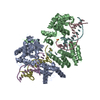
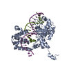
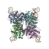
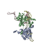
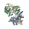
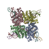
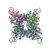
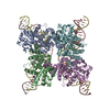
| Title | BASIS FOR A SWITCH IN SUBSTRATE SPECIFICITY: CRYSTAL STRUCTURE OF SELECTED VARIANT OF CRE SITE-SPECIFIC RECOMBINASE, LNSGG BOUND TO THE ENGINEERED RECOGNITION SITE LOXM7 | |||||||||
 Components Components |
| |||||||||
 Keywords Keywords | RECOMBINATION/DNA / CRE / Recombinase / DNA / CRE-LOXP RECOMBINATION / RECOMBINATION-DNA COMPLEX | |||||||||
| Function / homology |  Function and homology information Function and homology information | |||||||||
| Biological species |  Escherichia phage P1 (virus)Escherichia virus P1 Escherichia phage P1 (virus)Escherichia virus P1 | |||||||||
| Method |  X-RAY DIFFRACTION / SYNCHROTRON / FOURIER SYNTHESIS / Resolution: 2.75 Å X-RAY DIFFRACTION / SYNCHROTRON / FOURIER SYNTHESIS / Resolution: 2.75 Å | |||||||||
 Authors Authors | Baldwin, E.P. / Martin, S.S. / Abel, J. / Gelato, K.A. / Kim, H. / Schultz, P.G. / Santoro, S.W. | |||||||||
 Citation Citation | Journal: Chem.Biol. / Year: 2003 Title: A specificity switch in selected cre recombinase variants is mediated by macromolecular plasticity and water. Authors: Baldwin, E.P. / Martin, S.S. / Abel, J. / Gelato, K.A. / Kim, H. / Schultz, P.G. / Santoro, S.W. #1: Journal: Proc.Natl.Acad.Sci.USA / Year: 2002Title: Directed evolution of the site specificity of Cre recombinase. Authors: Santoro, S.W. / Schultz, P.G. #2: Journal: Biochemistry / Year: 2003Title: Modulation of the active complex assembly and turnover rate by protein-DNA interactions in Cre-LoxP recombination. Authors: Martin, S.S. / Chu, V.C. / Baldwin, E. #3: Journal: J.Mol.Biol. / Year: 2002Title: The order of strand exchanges in Cre-LoxP recombination and its basis suggested by the crystal structure of a Cre-LoxP Holliday junction complex. Authors: Martin, S.S. / Pulido, E. / Chu, V.C. / Lechner, T.S. / Baldwin, E.P. | |||||||||
| History |
|
- Structure visualization
| Structure viewer | Molecule: MolmilJmol/JSmol |
|---|
- Downloads & links
-Download
| PDBx/mmCIF format | 1pvq.cif.gz | 179.8 KB | Display | PDBx/mmCIF format |
|---|---|---|---|---|
| PDB format | pdb1pvq.ent.gz | 139.7 KB | Display | PDB format |
| PDBx/mmJSON format | 1pvq.json.gz | Tree view | PDBx/mmJSON format | |
| Others |  Other downloads Other downloads |
-Validation report
| Arichive directory | https://data.pdbj.org/pub/pdb/validation_reports/pv/1pvqftp://data.pdbj.org/pub/pdb/validation_reports/pv/1pvq | HTTPS FTP |
|---|
-Related structure data
| Related structure data |  1pvpC  1pvrC  1kbuS S: Starting model for refinement C: citing same article ( |
|---|---|
| Similar structure data |


-Links
 PDBj
PDBj


- Assembly
| Deposited unit | 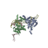
| ||||||||
|---|---|---|---|---|---|---|---|---|---|
| 1 | 
| ||||||||
| Unit cell |
| ||||||||
| Details | The second part of the tetrameric biological assembly is generated by the two fold axis: x, -y+1, -z+1. |
-Components
| #1: DNA chain | Mass: 10469.798 Da / Num. of mol.: 1 / Source method: obtained synthetically Details: TOP STRAND OF LOXM7 ENGINEERED DNA SUBSTRATE (LOXP(C7/G28,T8/A27,A9/T26) Source: (synth.) Escherichia virus P1 / References: GenBank: M10145 | ||
|---|---|---|---|
| #2: DNA chain | Mass: 10438.787 Da / Num. of mol.: 1 / Source method: obtained synthetically Details: BOTTOM STRAND OF LOXM7 ENGINEERED DNA SUBSTRATE (LOXP(C7/G28,T8/A27,A9/T26) Source: (synth.) Escherichia virus P1 / References: GenBank: M10494 | ||
| #3: Protein | Mass: 39222.805 Da / Num. of mol.: 2 / Mutation: I174L,T258N,R259S,E262G,E266G Source method: isolated from a genetically manipulated source Details: SELECTED CRE SITE-SPECIFIC RECOMBINASE MUTANT (I174L,T258N, R259S, E262G, E266G) Source: (gene. exp.) Escherichia phage P1 (virus) / Genus: P1-like viruses / Gene: cre / Plasmid: PET28B(+) / Species (production host): Escherichia coli / Production host:  #4: Water | ChemComp-HOH / |  Mass: 18.015 Da / Num. of mol.: 67 / Source method: isolated from a natural source / Formula: H2O Mass: 18.015 Da / Num. of mol.: 67 / Source method: isolated from a natural source / Formula: H2O |
-Experimental details
-Experiment
| Experiment | Method: X-RAY DIFFRACTION / Number of used crystals: 1 |
|---|
- Sample preparation
| Crystal | Density Matthews: 2.94 Å3/Da / Density % sol: 58.19 % | |||||||||||||||||||||||||||||||||||||||||||||||||||||||||||||||
|---|---|---|---|---|---|---|---|---|---|---|---|---|---|---|---|---|---|---|---|---|---|---|---|---|---|---|---|---|---|---|---|---|---|---|---|---|---|---|---|---|---|---|---|---|---|---|---|---|---|---|---|---|---|---|---|---|---|---|---|---|---|---|---|---|
| Crystal grow | Temperature: 294 K / Method: vapor diffusion, hanging drop / pH: 5.5 Details: Sodium Acetate, Calcium Chloride, 2,4-Methylpentanediol, pH 5.5, VAPOR DIFFUSION, HANGING DROP, temperature 294K | |||||||||||||||||||||||||||||||||||||||||||||||||||||||||||||||
| Components of the solutions |
| |||||||||||||||||||||||||||||||||||||||||||||||||||||||||||||||
| Crystal grow | *PLUS Temperature: 21-25 ℃ / Method: vapor diffusion / Details: Martin, S.S., (2002) J.Mol.Biol., 319, 107. / PH range low: 5.5 / PH range high: 5 | |||||||||||||||||||||||||||||||||||||||||||||||||||||||||||||||
| Components of the solutions | *PLUS
|
-Data collection
| Diffraction | Mean temperature: 100 K |
|---|---|
| Diffraction source | Source: SYNCHROTRON / Site: SSRL  / Beamline: BL7-1 / Wavelength: 1.08 Å / Beamline: BL7-1 / Wavelength: 1.08 Å |
| Detector | Type: MARRESEARCH / Detector: IMAGE PLATE / Date: Jun 12, 2002 |
| Radiation | Monochromator: DOUBLE-CRYSTAL / Protocol: SINGLE WAVELENGTH / Monochromatic (M) / Laue (L): M / Scattering type: x-ray |
| Radiation wavelength | Wavelength: 1.08 Å / Relative weight: 1 |
| Reflection | Resolution: 2.75→90 Å / Num. all: 31227 / Num. obs: 29853 / % possible obs: 95.6 % / Observed criterion σ(F): 0 / Observed criterion σ(I): -3 / Redundancy: 3.8 % / Rmerge(I) obs: 0.044 / Net I/σ(I): 17.3 |
| Reflection shell | Resolution: 2.75→2.85 Å / Rmerge(I) obs: 0.343 / Mean I/σ(I) obs: 3.8 / Num. unique all: 3001 / % possible all: 97.8 |
| Reflection | *PLUS % possible obs: 96 % |
| Reflection shell | *PLUS % possible obs: 98 % |
- Processing
| Software |
| ||||||||||||||||||||
|---|---|---|---|---|---|---|---|---|---|---|---|---|---|---|---|---|---|---|---|---|---|
| Refinement | Method to determine structure: FOURIER SYNTHESIS Starting model: PDB ENTRY 1KBU Resolution: 2.75→5 Å / Isotropic thermal model: Isotropic / Cross valid method: THROUGHOUT / σ(F): 0 / Stereochemistry target values: Engh & Huber
| ||||||||||||||||||||
| Refine analyze | Luzzati d res low obs: 5 Å / Luzzati sigma a obs: 0.27 Å | ||||||||||||||||||||
| Refinement step | Cycle: LAST / Resolution: 2.75→5 Å
| ||||||||||||||||||||
| Refine LS restraints |
| ||||||||||||||||||||
| Refinement | *PLUS Lowest resolution: 5 Å | ||||||||||||||||||||
| Solvent computation | *PLUS | ||||||||||||||||||||
| Displacement parameters | *PLUS |
